Conception and Development of Fertilized ovum
Subject: Midwifery I (Theory)

Overview
Conception, fertilization, or impregnation refers to the process of fusion or union of the spermatozoon with the mature ovum, which results in the fertilized single mono-nucleated cell known as the zygote. The ampullary section of the fallopian tube, which is the broadest and closest to the ovary, is the most common site of conception. Neither sperm nor ovum can survive for more than 2 or 3 days, and fertilization is most likely to occur when intercourse occurs within 48 hours of ovulation. The blastocyst's outer cell mass is derived from the trophoblastic layer. On the eighth day, the inner cell mass develops into the bilaminar germ disc, which is composed of a dorsal ectodermal layer of tall columnar cells and a ventral endodermal layer of flat polyhedral cells. During pregnancy, the endometrial lining of the uterus is referred to as decidua, and it is lost after delivery. During pregnancy, progesterone from the corpus luteum and placenta keeps decidua at bay.
Conception, fertilization, or impregnation refers to the process of fusion or union of the spermatozoon with the mature ovum, which results in the fertilized single mononucleated cell known as the zygote. The primary goals of fertilization are as follows:
- To stimulate egg embryonic development
- To restore the species' chromosomal complement
Normal Side of Conception
The ampullary component (Ampulla) of the fallopian tube, which is the widest region closest to the ovary, is the most common site of conception.
Usual Time for Conception
Neither sperm nor ovum can survive for more than 2 or 3 days, and fertilization is most likely when intercourse occurs within 48 hours of ovulation. As a result, conception will occur approximately 14 days before the next period is due. Sex chromosomes determine the sex of the new human at the time of conception. Every human cell includes 46 chromosomes, 44 conventional chromosomes, and two sex chromosomes. X and Y are the sex chromosomes. Women do not have a Y chromosome, although men do (male-44+X++Y) (female-44+X+X).
If 22+Y fertilizes the ovum, the Zygote will be 44+X+Y=male.
As a result, a child's sex is always determined by the father.
Development of Fertilized Ovum
Morula
Morula is an early-stage embryo composed of cells (called blastomeres) arranged in a solid ball within the zona pellucida.
A morula differs from a blastocyst in that it is a 16-cell mass in a spherical form, whereas a blastocyst (4–5 days post fertilization) has a cavity inside the zona pellucida as well as an inner cell mass. If left untreated and implanted, a morula will eventually grow into a blastocyst.
A series of cleavage divisions of the early embryo, beginning with the single-celled zygote, results in the morula. The embryo begins to resemble a mulberry once it has divided into 16 cells, hence the name morula (Latin, morus: mulberry). Cells on the outside of the morula become closely joined together with the creation of desmosomes and gap junctions within a few days following fertilization, becoming practically indistinguishable. This is referred to as compaction. A hollow formed from within the morula by the active transfer of sodium ions from trophoblast cells and water osmosis.
Blastocyst
About 5 days after fertilization, a fluid-filled cavity opens up in the morula, a ball of a few hundred cells. Following fast cleavage, the blastocyst has a diameter of roughly 0.1-0.2 mm and a cell count of 200-300. (cell division). The blastocyst begins to embed itself into the endometrium of the uterine wall after about 1 day (5–6 days post-fertilization), which is the time it generally takes to reach the uterus, where it will undergo later developmental processes, including gastrulation. The blastocyst must hatch from the zona pellucida in order to be embedded into the endometrium, which prevents it from adhering to the oviduct as it travels to the uterus. Only 11–12 days after fertilization, the blastocyst is entirely entrenched in the endometrium.
Implantation
The survival and development of the early embryo are dependent on implantation. It forges a bond between the mother and the early embryo that will last the duration of the pregnancy. Implantation is enabled by structural changes in both the blastocyst and the endometrial wall. Hatching occurs when the zona pellucida around the blastocyst breaches. This removes the physical limit on the growth of the embryonic mass and exposes the blastocyst's outer cells to the inside of the uterus. Furthermore, hormonal changes in the mother, notably an increase in luteinizing hormone (LH), prepare the endometrium to receive and encapsulate the blastocyst. Once attached to the endometrium's extracellular matrix, trophoblast cells produce enzymes and other substances that help the blastocyst get embedded in the uterine wall. The secreted enzymes damage the endometrial lining, while autocrine growth hormones including human chorionic gonadotropin (HCG) and insulin-like growth factor (IGF) allow the blastocyst to infiltrate the endometrium further.
Gastrulation, which includes the creation of the placenta from trophoblastic cells and differentiation of the ICM into the amniotic sac and epiblast, is enabled by implantation in the uterine wall.
The Early Development of the Embryo
The blastocyst's outer cell mass is derived from the trophoblastic layer. On the eighth day, the inner cell mass develops into the bilaminar germ disc, which consists of a dorsal ectodermal layer of tall columnar cells and a ventral endodermal layer of flat polyhedral cells. The bilaminar germs disc is linked to the trophoblast by the body stalk, a mesenchymal connecting stalk that eventually becomes the umbilical cord.
Formation of Amniotic Cavity and Yolk Sac
On either side of the bilaminar embryonic disc, two cavities emerge. The amniotic cavity is the dorsal cavity between the ectoderm and trophoblastic layers, which is bordered by mesenchyme. The yolk sac is a hollow on the ventral aspect that is lined on the outside by primitive mesenchyme and on the inside by the endodermal layer of the germ cell disc.
Germinal Layers and Their Derivatives
The three germ layers develop to generate the following tissues and organs:
- Ectodermal layer: Develops the central and peripheral nerve systems, skin and its appendages, the pituitary gland, the lining of the mouth, paranasal sinuses, and the roof of the mouth, among other things.
- Mesodermal layer: Develops bones, cartilage, muscles, the cardiovascular system, the kidneys, gonads, salivary glands, suprarenal glands, the spleen, the genital tract, the peritoneum, the pleura, and the pericardium.
- Endodermal layer: Develops gastrointestinal epithelial lining, liver, gall bladder, pancreas, respiratory epithelial lining, the mucous membrane lining the bladder and urethra, and larger vestibular glands.
Trophoblast
Small projections begin to form all over the blastocyst's surface, becoming most abundant at the point of contact. These trophoblastic cells divide into three distinct layers:
- The outer syncytiotrophoblast (syncytium) is made up of nucleated protoplasm that is capable of breaking down tissue during the embedding process. It erodes the decidua blood vessel walls, allowing nutrients in the maternal blood to reach the developing fetus.
- The inner cytotrophoblast is a distinct single layer of cells that produces the hormone human chorionic gonadotrophin (HCG). This is in charge of notifying the corpus luteum that pregnancy has begun. The corpus lutetium continues to produce estrogen and progesterone, which help to keep the decidua intact.
- A layer of mesoderm or primitive mesenchyme consists of loose connective tissue in the inner aspect. There is similar tissue in the inner cell mass and the two are continuous at the point where they join in the body stalk.
The trophoblast is responsible for the formation of the placenta and fetal membrane, which perform important functions such as attaching the fetus to maternal tissues, providing nutrition, oxygenation, clearing fetal metabolic wastes, and producing hormones, all of which contribute to the fetus's growth and development.
Decidua
During pregnancy, the endometrial lining of the uterus is referred to as decidua, and it is lost after birth. During pregnancy, progesterone from the corpus luteum and placenta maintains decidua. It is known as the decidual response.
A well-developed decidua has three layers:
- The superficial compact layer is made up of a dense mass of decidual cells, glands, ducts, and dilated capillaries. The majority of the surface epithelium is either thinned or gone.
- Dilated uterine glands, decidual cells, and blood arteries comprise the intermediate spongy layer (cavernous or functional layer). This layer is when the placenta separates.
- This basal layer is opposite to the uterine muscle and contains the basal component of the glands. Following parturition, the mucous coat regenerates from this layer.
Following the implantation of the blastocyst into the decidua's compact layer, the various parts of the decidua are renamed as follows:
- Decidua basalis or Serotina: The part of the decidua in contact with the blastocyst's base.
- Decidua Capsularies or Reflexa: The blastocyst's thin superficial compact layer.
- Decidua vera or Parietalis: The remaining decidua lining the uterine cavity outside of the implantation location.
Functions
- To make room for the fertilized ovum's implantation
- To supply nutrients (glycogen and lipids) to the developing ovum
- Provide a safeguarding action
- Provide the placental basal plate
References
- BabyCenter. November 2016. <http://www.babycentre.co.uk/1-week-conception>.
- Wong, D.L., 1995, Whaley and Wongs, Nursing care of infants and Children, 5th edition, Mosby, St.louis, London, Philadelphia
- Babycenter. Fetal development week by week Accessed 9/11/2014.
- Jones, D.L., 1973, Fundamentals of obstetrics and gynecology Volume 1, Obstetrics, reprint first edition, Derek Llewellyn Jone, 1969.
- WomensHealth.gov. Stages of pregnancy Accessed 9/11/2014.
- Tuitui R. 2002, A textbook of Midwifery A (Antenatal), 3rd edition, Vidyarthi Pustak Bhandari (Publisher and Distributor), Bhotahity, Kathmandu
Things to remember title
- Conception, fertilization, or impregnation refers to the process of fusion or union of the spermatozoon with the mature ovum, which results in the fertilized single mononucleated cell known as the zygote.
- The ampullary component (Ampulla) of the fallopian tube, which is the widest region closest to the ovary, is the most common site of conception.
- Neither sperm nor ovum can survive for more than 2 or 3 days, and fertilization is most likely when intercourse occurs within 48 hours of ovulation.
- Every human cell includes 46 chromosomes, 44 conventional chromosomes, and two sex chromosomes. The blastocyst's outer cell mass is derived from the trophoblastic layer.
- On the eighth day, the inner cell mass develops into the bilaminar germ disc, which consists of a dorsal ectodermal layer of tall columnar cells and a ventral endodermal layer of flat polyhedral cells.
- During pregnancy, the endometrial lining of the uterus is referred to as decidua, and it is lost after birth.
- During pregnancy, progesterone from the corpus luteum and placenta maintains decidua.
Videos for Conception and Development of Fertilized ovum
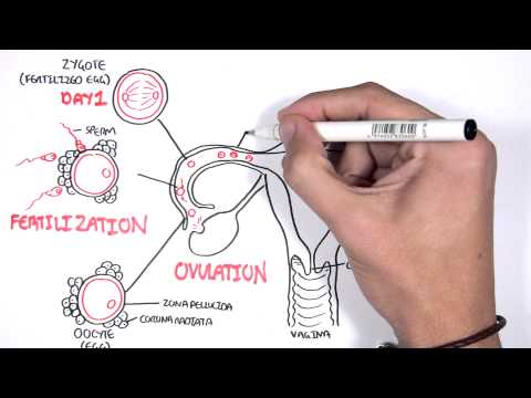
Embrology - Day 0 7 Fertilization, Zygote, Blastocyst
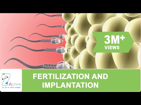
FERTILIZATION AND IMPLANTATION

The process of fertilization and the formation of human
Questions and Answers
What do you mean by conception, write its normal site and usual timing of conception?
Conception, fertilization, or impregnantation are terms used to describe the process of the spermatozoon joining with the mature ovum to form the fertilized zygote, a single mononucleated cell. Fertilization's primary goals are:
- To start the egg's embryonic development.
- To make the species' chromosome complement whole.
Normal side of conception:
The ampullary portion (Ampulla), which is the widest portion closest to an ovary, of the fallopian tube, is where conception most frequently occurs.
Usual time for conception:
Because neither sperm nor ovum can survive for more than two or three days, fertilization is most likely to happen when sexual activity occurs within 48 hours of ovulation. Therefore, it follows that conception will occur roughly 14 days before the start of the following period. Sex chromosomes control the sex of the unborn child at the time of conception. Every human cell has 46 chromosomes, consisting of 2sex chromosomes and 44 regular chromosomes. There are two sex chromosomes: X and Y. Males have Y chromosomes (male-44+X++Y) while females lack Y chromosomes (female-44+X+X).
If 22+Y fertilizes the ovum, the resulting zygote will be 44+X+Y=male.
As a result, a child's gender is always decided by the father.
Explain the development of fertilized ovum.
Development of fertilized ovum
- Morula
A morula is a young embryo made up of cells (known as blastomeres) arranged in a solid ball inside the zona pellucida. The difference between a morula and a blastocyst is that a morula (3–4 days after fertilization) is a 16–cell mass that is shaped like a sphere, whereas a blastocyst (4-5 days after fertilization) has an inner cell mass and a cavity inside the zona pellucida. A morula will eventually transform into a blastocyst if left untreated and allowed to stay implanted.
A series of cleavage divisions of the early embryo, beginning with the single-celled zygote, results in the morula. The embryo is called a "morula" once it has divided into 16 cells and starts to resemble a mulberry (Latin, morus: mulberry). Within a few days of fertilization, desmosomes and gap junctions are formed, tightly binding the outermost portion of the morula's cells together and making them nearly indistinguishable. Compaction is the term for this action. Through the active transport of sodium ions from trophoblast cells and water osmosis, a cavity develops inside the morula.
- Blastocyst
About 5 days after conception, the morula, a ball of a few dozen cells, begins to form a fluid-filled chamber, signaling the start of blastocyst development. Following fast cleavage, the blastocyst has a diameter of 0.1–0.2 mm and contains 200–300 cells (cell division). The blastocyst usually takes about one day to reach the uterus (5–6 days post-fertilization), after which it starts to embed itself in the endometrium of the uterine wall where it will go through later developmental stages, including gastrulation. The blastocyst must emerge from the zona pellucida in order to be embedded into the endometrium; this prevents it from adhering to the oviduct as it travels to the uterus. Only 11–12 days after fertilization is the blastocyst fully embedded in the endometrium.
- Implantation
The early embryo's survival and growth depend on implantation. It forges a bond between the mother and the developing embryo that will last for the duration of the pregnancy. The blastocyst and endometrial wall undergo structural changes to enable implantation. It is known as hatching when the zona pellucida surrounding the blastocyst breaks. This releases the restriction on the embryonic mass's physical size and allows the blastocyst's outer cells to contact the uterine cavity.Furthermore, the endometrium is ready to receive the blastocyst and surround it thanks to hormonal changes in the mother, particularly a rise in luteinizing hormone (LH). When the blastocyst is attached to the endometrium's extracellular matrix, trophoblast cells secrete enzymes and other substances that help the blastocyst get embedded in the uterine wall. The endometrial lining is degraded by the enzymes released, and the blastocyst can further invade the endometrium thanks to autocrine growth factors like human chorionic gonadotropin (HCG) and insulin-like growth factor (IGF). The following stage of embryogenesis, known as gastrulation, which involves the formation of the placenta from trophoblastic cells and the differentiation of the ICM into the amniotic sac and epiblast, is made possible by implantation in the uterine wall.
- The early development of the Embryo
The trophoblastic layer's blastocyst's outer cell mass. On the eighth day, the inner cell mass develops into the bilaminar germ disc, which consists of a ventral endodermal layer made up of flat polyhedral cells and a dorsal ectodermal layer of tall columnar cells. The body stalk, a mesenchymal connecting stalk that later takes the form of the umbilical cord, connects the bilaminar germs disc to the trophoblast. Formation of the yolk sac and amniotic cavity. There are two chambers on either side of the bilaminar embryonic disc. The term "amniotic cavity" refers to the dorsal cavity between the ectoderm and trophoblastic layers that is lined by mesenchyme. The yolk sac is the term for the cavity on the ventral aspect that is lined on the outside by primitive mesenchyme and inside by the endodermal layer of the germ cell disc.
The following list of tissues and organs are formed from the three germ layers through differentiation:
- Ectodermal layer: Develops the pituitary gland, the central and peripheral nervous systems, the skin and its appendages, the paranasal sinuses, the roof of the mouth, and other structures.
- Mesodermal layer: Develops the skeleton, cartilage, muscles, peritoneum, pleura, pericardium, kidneys, gonads, suprarenal glands, spleen, genital tract, and cardiovascular system.
- Endodermal layer: Develops the mucous membrane lining the bladder and urethra, the greater vestibular glands, the respiratory tract, the liver, the gall bladder, the pancreas, the gastrointestinal system, and the respiratory epithelial lining.
All over the blastocyst's surface, small projections start to appear, but they are most prevalent where there is contact. Three layers of differentiation occur in these trophoblastic cells:
- The nucleated protoplasm that makes up the outer syncytiotrophoblast (syncytium) is capable of dissolving tissue after embedding. It erodes the decidua's blood vessel walls, making nutrients in the mother's blood available to the growing fetus.
- A distinct single layer of cells called the inner cytotrophoblast produces the hormone known as human chorionic gonadotrophin (HCG). This is what alerts the corpus luteum that a pregnancy has started. Estrogen and progesterone are still produced by the corpus luetum, which helps to keep the decidua healthy.
- Mesoderm, or primitive mesenchyme, a layer of loose connective tissue, is present in the inner aspect. The inner cell mass contains tissue that is similar, and the two are continuous where they meet in the body stalk.
The trophoblast is in charge of creating the placenta and fetal membrane, which perform crucial tasks like securing the fetus to the mother's tissues, supplying nutrition, oxygenation, and clearing away metabolic wastes while also producing hormones to promote the fetus' growth and development.
- Decidua
During pregnancy, the uterus' endometrial lining is referred to as decidua, and it sheds after delivery. During pregnancy, progesterone produced by the placenta and corpus luteum keeps the decidua in place. The reaction is known as the decidual one.
Three layers make up the decidua when it is fully developed:
- Superficial compact layer: Consists of a dense mass of capillaries, glands, ducts, and decidual cells. The majority of the surface epithelium either thins out or disappears.
- Intermediate spongy layer (cavernous or functional layer): Consists of blood vessels, blood cells, and dilated uterine glands. In this layer, placenta separation takes place. This basal layer, which is opposite the uterine muscle, contains the basal portion of the glands. Following parturition, the mucous coat regenerates from this layer.
The various parts of the decidua are given new names after the blastocyst is implanted into the compact layer of the decidua:
- Decidua basalis or serotina: The area of the decidua that comes into contact with the blastocyst's base.
- Decidua capsularies or reflexa: The blastocyst's thin, superficial compact layer.
- Decidua vera or parietalis: Outside of the site of implantation, the remainder of the decidua lines the uterine cavity.
The function of the decidua are:
- To create a location for the fertilized ovum to be implanted.
- Give the developing ovum nutrition (glycogen, fat)
- Take a protective measure
- Give the placenta's basal plate.
© 2021 Saralmind. All Rights Reserved.
 Login with google