The structure and function of tissue: Epithelial tissue, connective tissue, muscular tissue, nervous tissue-1
Subject: Anatomy and Physiology

Overview
Tissues
Highly ordered units make up cells. However, they don't work alone. They collaborate in tissues, which are collections of related cells. A tissue is a collection of comparable cells and the intercellular material they are made of that has a comparable embryological origin and works as a unit to carry out a specific function. Histology is a science that focuses on the investigation of a tissue.
According to their structure and function, the body's various tissues are divided into four main categories.
- Epithelial tissue
- Connective tissue
- Muscular tissue
- Nervous tissue
Epithelial Tissue
Cells are organized in single or many layers in continuous sheets to form epithelial tissue. They can be found as the linings of the body's ducts, glands, and internal body surfaces (inner surfaces). They always have a free surface since they are never covered by another tissue. Despite not having its own blood supply, an epithelial tissue possesses its own nerve supply. The blood vessels in the nearby connective tissue transport nutrients in and waste out. The epithelial tissues' various functions include defense, filtration, absorption, secretion, and excretion. They fall into two categories:
- Covering and lining epithelium
- Glandular epithelium
Covering and Lining Epithelium
- It creates the outer layer that protects some internal organs as well as the external body surface. It comprises the nerve tissue, the lining of the body cavity, the inside of the gastrointestinal and respiratory tracts, and blood vessels (the parts of sense organs for smell, hearing, touch). Gametes (egg and sperm) develop from this tissue. Based on the arrangement of cells in layers and cell shapes, covering and are categorized.
Covering and lining epithelium are categorized as follows depending on how the layers are arranged:
- Simple Epithelium
- It is made up of just one layer of cells and is designed for wear-free secretion, absorption, diffusion, and filtration.
- Stratified Epithelium
- It has two or more layers of cells and is located in an area that experiences a lot of wear and tear.
- Pseudo-Stratified Epithelium
- It is a unique variety of simple epithelium made up of a single layer of erroneously stratified epithelial cells resting on a basement membrane. It appears to have several cell layers.
Covering and lining epithelium are categorized into the following:
- Squamous
- Cells are flattened, like floor tiles and are thin.
- Cuboidal
- Tall and cube-like cells are present.
- Columnar
- The cylindrical and significantly taller cells.
- Transitional
- Combination of cell shapes, such as cuboidal to columnar, cuboidal to polyhydral, and cuboidal to squamous, that are present in areas of significant distention or expansion. The urinary bladder is one example.
Simple Epithelium
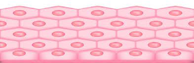
- Simple-Squamous Epithelium
- Consists of a single layer of flat cells that give the impression of a tiled floor. The air sac, blood vessels, and lymph vessels are lined with it.
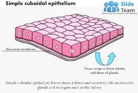
- Simple-Cuboidal Epithelium
- Contains flat polygonal cells that line the thyroid gland, the anterior surface of the eye's lens, the retina, and the surface of the ovary.
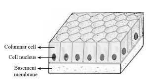
- Simple-Columnar Epithelium
- Simple columnar epithelium has cells that resemble columns (taller than they are wide). Various modifications are made depending on the place and purpose. It lines the gastrointestinal tract, the gall bladder, and several glands' excretory tubes. It performs activities in lubrication, secretions, absorption, and protection.
- Pseudo Stratified Columnar (ciliated and non-ciliated epithelium)
- Cilia are the tiny hair-like processes of columnar cells. Microtubules enclosed in a plasma membrane, which protrudes from the columnar cells' free edge, make up the cilia. The contents of the tube are propelled by the cilia's wave-like movement. They are located in the respiratory system and uterine tube lining. Cells are present in the pseudo-stratified columnar epithelium of the non-ciliated type. without cilia and their ability to absorb and protect.
Stratified Epithelium
It is more resilient and shields underlying tissues from the outside environment and from deterioration.
- Stratified Squamous Epithelium
- The outer cells of this form of epithelium are flat. Based on the presence of keratin, stratified squamous epithelium is categorized into two groups. Which are: Squamous epithelium that is stratified and keratinized. It is common to find non-keratinized stratified squamous epithelium on wet surfaces that have experienced significant wear and tear.
- For instance, the mouth, tongue, and vagina. The surface cell of this kind creates a strong layer of keratin-containing material in keratinized stratified squamous epithelium. Consider skin. Keratin, a protein that is 15 waterproof, fends off bacterial invasion and friction.
- Stratified Cuboidal Epithelium
- It is an uncommon kind of epithelium. It can be found in esophageal glands, cavernous urethras, and sweat glands. Its primary purposes are absorption and protective limited secretion.
- Stratified Columnar Epithelium
- It is also unusual for the body. The anus layers and milk duct of the mammary gland contain stratified column epithelium. It performs secretion and defense functions.
- Transitional Epithelium
- With the exception of the fact that the cells in the apical layer typically tend to be large and rounded, it resembles stratified cuboidal epithelium. This epithelium's elasticity makes it perfect for structures like the urinary bladder that are prone to internal expansion.
Pseudo Stratified Epithelium
All of the cells in the single layer lie on the basement membrane, but none of them reach the apical surface, giving the appearance that the layer is stratified. In order to give the illusion of stratification, the nuclei, which are normally the most noticeable cellular structure, are widely spaced apart. These characteristics provide the appearance of a multilayered tissue when viewed from the side, earning the label pseudo stratified epithelium. The human body contains two primary subtypes of pseudostratified columnar epithelia:
The upper respiratory tract, particularly the trachea, is lined by respiratory epithelium, a ciliated pseudostratified columnar epithelium. The male reproductive system's epididymis is lined by a non-ciliated pseudostratified columnar epithelium.
Glandular Epithelium
Secretion is their primary function. Glands fall into one of two categories:
- Excocrine
- Those glands that secrete into tubes or ducts that terminate at the surfa covering. Mucus, oil, wax, perspiration, and digestive enzymes are their main byproducts. Exocrine glands include salivary and sweat glands.
- Endocrine
- In the end, they secrete their goods into the bloodstream. Hormones are constantly present in an endocrine gland's secretions. Chemicals called hormones control a number of physiological processes. Endocrine organs include pituitary, thyroid, and adrenal glands.
Classification of Exocrine Glands
They are categorized based on how they are built and how the secretary portion is shaped. They can be classified into the following groups:
- Unicellular Gland
- Possess a single cell. The respiratory, gastrointestinal, and genitourinary systems' goblet cells are the best examples.
- Multicellular Gland
- Are made up of numerous cells and come in a variety of shapes. Salivary and sebaceous glands are examples.
Exocrine glands can be categorized by looking at the secretary portion into:
- Tubular Gland
- If a gland's secretory portion is tubular.
- Acinar Gland
- If the secretary part resembles a flask.
- Tubulo-Acinar
- If it has both a flask- and a tubular-shaped secretary portion.
In addition, a gland is referred to as a simple gland if its duct does not branch, and a compound gland if it does. Exocrine glands are divided into the following categories by combining the shape of the secretary portion with the degree of duct branching:
- Unicellular
- Multi-cellular
- Simple tubular
- Compound tubular
- Simple coiled tubular
- Compound acinar
- Simple acinar
- Compound tubulo -acinar
- Simple branched acinar
- Tubulo-acinar
Things to remember title
© 2021 Saralmind. All Rights Reserved.
 Login with google