Circulatory system: Structure and function of heart
Subject: Anatomy and Physiology

Overview
The circulatory system is the body's transportation system, carrying nutrients, oxygen, water, and other necessities to tissue cells as well as removing waste from those cells. It is composed of three sections:
- The blood:
- The fluid that carries materials to and from the tissue is called blood.
- The blood vessels:
- The pathways via which the blood enters, passes through, and returns to the heart
- The heart:
- The driving force behind the blood's motion is the heart.
Cardiology is the scientific study of the healthy heart and the internal disorders that affect it.
The Heart Structure
The size of a closed fist would closely describe the heart, a tiny muscular organ. An adult heart measures around 12 cm in length, 9 cm at the base, and 6 cm in thickness. It weighs roughly 300g for men and 250g for women.
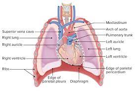
Location of the Heart
The mediastinum, or space between the lungs, is where the heart is housed in the thoracic cavity. It is between the second and sixth ribs (2nd -6th ). It is located to the left of the body's midline by about two thirds. The place of attachment for the major vessels is the base, a large region on top of the heart. The apex, which is its inferior end, rests on the diaphragm and teeters to the left.
Organs Associated with the Heart (relation of heart)
- Superiorly-
- The great blood vessels, i.e. the aorta, superior vena cava, pulmonary artery and pulmonary vein
- Inferiorly-
- The apex rests on the central tendon of the diaphragm
- Anteriorly-
- The sternum, ribs and intercostal muscles
- Posteriorly-
- The oesophagus, trachea, left and right bronchus, descending aorta, inferior vena cava and thoracic vertebrae Laterally- the lungs- left lung overlaps the left side of the heart
The Pericardium
The pericardium, which is connected to the connective tissue of the major vessels above the heart and to the diaphragm below, is a double-walled sac that surrounds the heart.
In the pericardium, there are two layers:
- Fibrous pericardium:
- A stiff, elastic, thick, and uneven connective tissue makes up the outer fibrous pericardium. The heart is shielded from overstretching by the fibrous pericardium.
- The pericardial inner serous layer:
- It is a thin, smooth layer that is made up of two layers. The serous pericardium's outer parietal layer and the fibrous pericardium are fused together. The epicardium is another name for the serous pericardium's inner visceral layer. One of the layers of the heart wall is the epicardium.
A region known as the pericardial cavity is located between the two layers of serous pericardium. Pericardial fluid, a thin lubricating fluid, can make up to 50 ml of it. The membranes are lubricated by this fluid, which also permits the heart to beat without resistance.
Layers of the Heart wall
Three layers make up the heart wall:
The Epicardium
The heart wall's outermost, thinnest layer is called the epicardium. Simple squamous epithelium and delicate connective tissue make up its structure. The visible layer of the serous pericardium is another name for the epicardium.
The Myocardium
The middle layer of muscle in the heart, known as the myocardium, is what pumps blood. The heart muscle that makes up this thickest layer is involuntary and striated like skeletal muscle. The branched, short, dense cells that make up the heart muscle are called cardioblasts. Thick intercalated discs form a link between the ends of nearby cells in the heart muscle. Synchronized contractions are made possible by this link, which enables direct impulse transmission throughout the entire heart. Because it pumps more blood throughout, the left ventricle has the thickest myocardium.
The Endocardium
The innermost layer, the endocardium, is made up of a straightforward squamous endothelium that sits on top of a thin layer of connective tissue. It covers the heart valves and offers a smooth lining for the heart chambers. It blends seamlessly with the big blood arteries that are connected to the heart's endothelium lining.
Internal Structures of Heart
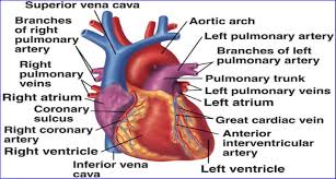
The Chambers of the Heart
Four chambers make up the heart. The two inferior pumping chambers are called ventricles, while the two superior receiving chambers are called atria.
- The Atria:
- The interatrial septum is a wall that separates the two atria from one another. Through the atrioventricular valves, the atria pump blood into the ventricles below. Three veins—the coronary sinus, inferior venacava, and superior venacava—supply blood to the right atrium. Four pulmonary veins supply blood to the left atrium from the lungs.
- The Ventricles:
- The interventricular septum is a substantial wall that divides the right ventricle from the left ventricle. The right ventricle's wall is only moderately thick and muscular because it primarily pumps blood to the back and lungs. The thickest cardiac chamber is the left ventricle. Blood is pumped through the entire body by this chamber, which has the heaviest workload of the four. The cusps of the atrioventricular valve are attached to papillary muscles on the ventricle floor by chordae tendinae (cords that resemble tendon), which are found in the internal walls of the heart.
- The Valves of the Heart:
- The heart needs valves that guarantee a one-way flow of blood in order to successfully pump blood. Each atrial has a valve that connects it to its ventricle at the point when the ventricle leaves the body and enters the great artery. Each valve is made up of two or three cusps, which are endothelium-coated fibrous flaps of tissue.
- The Atrioventricular Valves:
- Between an atrium and a ventricle are the atrioventricular (AV) valves. Because to its three cusps, the right AV valve is also known as the tricuspid valve, and the left AV valve is known as the bicuspid valve due to its two cusps. The mitral valve is another name for the left AV valve. The chordae tendineae that connect the AV valve cusps to the conical papillary muscles resemble a string.
- The AV valve cusps hang down limply, both AV valves are open, and blood readily flows from the atria into the ventricles when the ventricles are relaxed. Blood pressure causes the cusps to rise until their edges touch as the ventricles contract after filling with blood. This closes the apertures and stops blood from returning to the atria. The papillary muscle contracts a pulls the chordae tendineae, which prevents the valves from bulging (prolapsing) into the artia.
- The Semi lunar Valves:
- The pulmonary and aortic valves, which control the flow of blood from the ventricles into the major arteries, are part of the semi-lunar (SL) valves. The opening from the right ventricle into the pulmonary trunk is controlled by the pulmonary valve, and the opening from the left ventricle into the aorta is controlled by the aortic valve. It features three cusps on each valve.
- The chambers experience pressure buildup when the ventricles constrict. When the pressure in the ventricles is greater than the pressure in the arteries, the semi-lunar valves open, allowing blood to be ejected from the ventricles into the pulmonary artery and aorta. Arterial blood temporarily flows back toward the heart as the ventricles relax, filling the semi-lunar valve cusps. This causes the semi lunar valves to close thereby preventing blood from re-entering the heart.
Blood Flow through the Heart:
The pulmonary circulation and systemic circulation are two closed pathways through which the heart pumps blood after birth. In contrast to the left side of the heart, which pumps oxygenated blood into the systemic circulation, the right side of the heart expels blood into the pulmonary circulation after receiving deoxygenated (O2 deficient) blood. the left side of the heart is the recipient of blood returning from the pulmonary circulation.
Although blood is constantly bathing the heart chambers, the heart tissue receives very little nutrition from this blood. A network of arteries and capillaries within the heart carries blood to all areas of the cardiac circulation. Despite making up only 0.5% of the body's weight, the heart uses 5% of the blood in circulation to meet its own metabolic requirements.
Things to remember title
© 2021 Saralmind. All Rights Reserved.
 Login with google