circulatory system: Conduction system of heart and Cardiac cycle
Subject: Anatomy and Physiology

Overview
Blood Supply
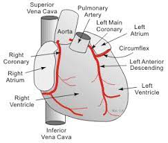
The arterial blood supply of the heart is provided by the right and left coronary arteries. These arteries branch from the ascending aorta immediately after the aorta leaves the left ventricles. The coronary arteries along with their extensive branches supply oxygenated blood to the myocardium. At rest, these vessels supply the myocardium with about 250 ml of the blood per minute.
Venous Drainage
Venous drainage refers to the route by which blood leaves an organ. After flowing through capillaries of the myocardium, about 20% of the coronary blood empties directly from small veins into the right ventricle. The other 80% returns to the right atrium by an enlarged vessel called the coronary sinus which is formed by union of several small veins.
Nerve Supply
The heart beats continuously due to its own intrinsic impulses which will be described shortly. In addition to it, the sympathetic and parasympathetic (vagus) nerves supply the heart. The sympathetic stimulation increases the rate and the force of the heart beat. The parasympathetic nerve supply decreases the rate and force of the heartbeat.
Heart Physiology (Function)
Cardiac Conduction System
The cardiac muscle has the ability to generate rhythmic electrical activity which allows the heart to beat continuously. It is a property of the heart muscle itself and does not depend on extrinsic nerve impulse. These self excitable muscle cells stimulate a heart to beat even after it is removed from the body.
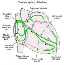
The network of specialized cardiac muscle cells which initiate and distribute impulses throughout the heart is known as the cardiac conduction system.It controls the route and timing of the electrical conduction to ensure that four chambers are co-ordinated with each other. The conduction system consists of the following components:
- Sinoatrial node (SA node).
- Atrioventricular node (AV node).
- Atrioventricular bundle (AV bundle).
- Purkinje fibers Sinoatrial Node (SA node.)
Sinoatrial Node (SA Node): This is a small patch of specialized cell located in the superior posterolateral wall of right atrium immediately below and slightly lateral opening of superior vena cava. The SA node is also known as the pacemaker of heart because it initiates each heart beat and sets the pace for the heart as a whole. Signals from the SA node spread throughout the atria causing atrial contraction. SA node typically depolarizes spontaneously at the rate of 60 to 80 times every minute.
The Atrioventricular Node (AV Node): The AV node is located in the inferior portion of the inter-arterial septum near the tricuspid valve. This node acts as an electrical gateway to the ventricles. The fibrous ring that separates atria and ventricles insulates and prevent currents from getting to ventricles by any other route. After spreading throughout the atria, the electrical impulses reach and stimulate the AV node. The AV node too can initiate impulses but at a slower rate than the SA node. The AV node initiates the impulses at the rate of 40-60 time/min. If SA node becomes inactive, the AV nodal rate will then determine the ventricular rate.
The Atrioventricular Bundle (AV Bundle): The AV bundle is a pathway by which electrical impulses leave the AV node. The AV bundle is also known as bundle of his. It is divided into right and left bundle branches that extend through the inter-ventricular septum and descend toward the apex of the heart. The electrical impulses then pass along both right and left bundle branches. It also initiates impulse at the rate of 25-40 beats/min.
Purkinje Fibers: These are nerve like processes that arise from the bundle branches near the apex of the heart. They then turn upward and spread throughout the ventricular myocardium. Purkinje fibers distribute the electrical excitation to the ventricular myocardium. Then the ventricles contract and push the blood upward towards the pulmonary artery and the aorta. It too initiates the rate of 25-40 beat/min.
The Cardiac Cycle
As we know that the heart pumps blood throughout the body continuously. For this activity, it contracts, forces blood out of its chamber and then relaxes allowing its chambers to refill with blood. The terms systole and diastole refers respectively to these contraction and relaxation. This series of events associated with the flow of blood through the heart during one complete heart beat as known as the cardiac cycle. Thus, a cardiac cycle systole and diastole of the atria plus systole and diastole of the ventricles.
Phases of Cardiac cycle
When the heart rate is 75 beats/min, a cardiac cycle lasts 0.8 sec. This circular chain of events consists of:
- Atrial systole.
- Ventricular systole.
- Complete cardiac diastole (Relaxation period).
Atrial Systole
It is also known as rapid filling phase or presystole. During atrial systole, the atria are contracting. At the same time ventricles are relaxed. It lasts about 0.1 sec.
The SA node fires and the atria depolarize which causes atrial systole. As the atria contract,they exert pressure on the blood within their chambers. This propels the additional blood through open AV valves into the ventricles. Then the atria relax.
Ventricular Systole
As the atria go into diastole, the ventricles begin their contraction phase. This phase lasts about a 0.3 sec. This has following events:
- Isometric contraction period.
- Ejection period Isometric contraction period.
Isometric contraction period is the type of muscular contraction characterized by increase in tension, without any change in the length of muscle fibers. Isometric contraction of ventricular muscle is also called isovolumetric contraction. Immediately after atrial systole, the atrioventricular valves are closed due to increase in ventricular pressure, closure of atrioventricular valve produce 1st heart sound. Semilunar valves are already closed. Now, ventricles contract as closed cavities, in such a way that there is no change in the volume of ventricular chambers or in the length of muscle fibers. Only the tension increases in ventricular musculature. Because of increased tension in ventricular musculature during isometric contraction, the pressure increases sharply inside the ventricles. Thus, the pressure rise in ventricle, caused by isometric contraction is responsible for the opening of semilunar valves, leading to ejection of blood from the ventricles into aorta and pulmonary artery.
Ejection period: Due to the opening of semilunar valves and isotonic contraction of ventricles, blood is ejected out of both the ventricles. Hence, this period is called eiection period. It has two stages: rapid ejection and slow ejection stage.
Relaxation Period: During the relaxation period, the atria and the ventricles are both relaxed. This phase about 0.4sec.
Following events occur in this period:
- Protodiastole,
- Isometric relaxation period,
- Rapid filling phase,
- Slow filling phase,
- Last rapid filling phase.
Protodiastole: Protodiastole is the first stage of ventricular diastole, hence the name prodiastole. Due to the ejection of blood, the pressure in aorta and pulmonary artery increases pressure in ventricles drops. When intraventricular pressure becomes less than the pressure in aorta and pulmonary artery, the semilunar valves close (produce second heart sound). Atrioventricular valves are already closed. No other change occurs in the heart during this period. Thus, protodiastole indicates only the end of systole and beginning of diastole.
Isometric Relaxation Period: Isometric relaxation is the type of muscular relaxation, characterized by decrease in tension without any change in the length of muscle fibers. Isometric relaxation of ventricular muscle is also called Isovolumetric relaxation. During isometric relaxation period, once again all the valves of the heart are closed. Now, both the ventricles relax as closed cavities without any change in volume or length of the muscle fiber. Intraventricular pressure decreases during this period. Thus, the fall in pressure in the ventricles, caused by isometric relaxation is responsible for the opening of atrioventricular valves, resulting in filling of ventricles.
Rapid Filling Period: When atrioventricular valves are opened, there is a sudden rush of blood (which is accumulated in atria during atrial diastole) from atria into ventricles. So, this period is called first rapid filling period. Ventricles also relax isotonically. About 70% of filling takes place during this phase.
Slow Filling Phase: After the sudden rush of blood, the ventricular filling becomes slow. Now, it is called the slow filling. It is also called diastasis. About 20% of filling occurs in this phase.
Last Rapid Filling Phase: This phase occurs because of atrial systole. After slow filling period, the atria contract and push a small amount of blood into ventricles. About 10% of ventricular filling takes place during this period. Flow of additional amount of blood into ventricle due to atrial systole is called atrial kick.
Things to remember title
© 2021 Saralmind. All Rights Reserved.
 Login with google