IMMUNOLOGY
Subject: Basic Science Applied to Nursing

Overview
Introduction to Immunology
Immunology is the study of specific resistance to further infection by a particular microorganism or its products. It is the science which deals with the response of body against antigen. It is a very broad scientific discipline which is important for the most fields of medicine. Immunological mechanisms are involved in the protection of body against infectious agents.
Cells and Organs of Immune System
The tissues and organs of vertebrates contain a diverse variety of immune response-producing cells. In primary (central) lymphoid organs, the lymphoid stem cells become two major populations of T and B lymphocytes (eg, thymus, bone marrow). Both types of lymphocytes circulate throughout the body and move to secondary (peripheral) lymphoid organs after maturing in main lymphoid organs (eg, spleen, lymph nodes etc.).
Primary (Central) lymphoid Organs
Thymus:
Vertebrate tissues and organs are home to a wide range of immune response-producing cells. The lymphoid stem cells differentiate into two main populations of T and B lymphocytes in primary (central) lymphoid organs (eg, thymus, bone marrow). Both types of lymphocytes circulate throughout the body and mature in primary lymphoid organs before moving to secondary (peripheral) lymphoid organs (eg, spleen, lymph nodes etc.).
Only the long-lived fraction (5%) of these proliferating lymphocytes contributes the majority of the cells that move to the secondary lymphoid organs during fetal & early postnatal life, and probably for longer duration. After leaving the thymus, these immunologically capable T cells circulate through the blood and lymphatics, making about 75% of the human body's circulating lymphocytes. Immunologically capable T-lymphocytes are thought to have a substantially longer life span than B-lymphocytes, lasting months or even years (days or weeks).
Bone marrow:
Within the B-bone marrow, some lymphoid stem cells mature into immunoglobulin (Ig)-producing lymphocytes known as lymphocytes. In the early stages of their development, B-cells display IgM on their surface.
The B cells exhibit either surface IgM alone or in conjunction with IgA or IgG at the following stage of development. Then, in addition to C3 (complement 3), and Fc receptor (receptor for the Fc part of immunoglobulin), IgD is also added to the surface of B-cells, designating the B-cell as mature.
The cells are now developing receptors for hormones and immunoregulatory substances.
Secondary (peripheral) lymphoid organs
Lymph Node:
The lymph nodes are around the size of a bean and are located along lymphatic channel pathways. The paracortical zone, which is located between the cortical follicles and the base of the medullary cords, is organized into an outer cortex, an inner medulla, and a mature lymph node. Blood cells in circulation enter lymph nodes through specialized venules.
The T and B lymphocytes are largely separated into different anatomical compartments of lymph node:
- B-cell areas: the cortical follicles contain B-cells
- T-cell areas: the paracortical area contains T-lymphocytes and constitute thymus dependent area
Both types of lymphocytes migrate to sinusoidal spaces of medulla and pass into blood via efferent lymphatic vessels.
Spleen:
The spleen consists of a loosely organized medulla and a cortex made up of tightly packed B and T cells. The medulla is exterior to the cortex, unlike the lymph node. The presence of a significant number of B-cells makes the spleen a key location for the generation of antibodies.
Like lymph nodes, T and B cell areas are segregated:
- B-cell area: Perifollicular region, germinal center and marginal zone
- T-cell area : Thymus dependent area includes the lymphatic sheath and lies adjacent to the central arterioles.
Mucosa Associated Lymphoid Tissue (MALT):
The mucosa of the digestive, respiratory, and genitourinary systems, which are continually exposed to a variety of antigens, are immunologically protected by subepithelial accumulations of lymphoid tissue.
In the lungs, the lymphoid tissue can either be found as an evenly distributed collection of lymphocytes, plasma cells, and macrophages or as specialized aggregates with well-developed follicles (lingual, palatine, pharyngeal tonsils, appendix). Mucosa-associated lymphoid tissue is the name given to them (MALT). B and T lymphocytes, as well as phagocytes, are present in MALT. It assembles an independent, interconnected secretory system made up of cells that produce IgA or IgE.
Defense Mechanism
The defense mechanism in human body is divided into two types:
a. Nonspecific defense mechanism:
It involves two defense mechanisms
- Primary Defenses (First line defense mechanism):
- The main factors in the first line of defense against infection are mechanical, accompanied by some humoral and cellular factors.
- These defenses represent an attempt to prevent microorganisms from colonizing on skin and mucous membrane.
- Secondary Defenses (Second line defense mechanism):
- The second line of defense consists of humoral and cellular factors in the blood and tissues. The most important is phagocytic cells.
- Phagocytic cells polymorphonuclear are leukocytes (microphages), macrophages. Both microphages and macrophages are capable of amoeboid motility and chemotactic migration.
The important mechanisms in nonspecific defenses against infection are-
- Mechanical factors:
- Anatomical structure (skin, mucosa) Mucus secretion & mucus flow from mucosa
- Movement of the ciliated epithelium in the lower respiratory tract
- Peristalsis of gastrointestinal tract Urine flow in urinary tract
- Humoral factors:
- Lactic acid from sweat gland
- Hydrochloric acid in the stomach
- Unsaturated fatty acids secreted by the sebaceous gland
- Lysozyme in saliva and tear fluid
- Serum proteins such as C-reactive protein, haptoglobin, fibrinogen, transferring
- Antiviral interferon etc.
- Cellular factors:
- Normal flora of skin and mucosa
- Natural killer cells
- Professional phagocytes (Macrophages, microphages)
b. Specific Defense Mechanism
When the immune system is activated by microbial antigens, particular defense is obtained via antibodies and selectively reactive T cells. Cell-mediated immunity is attained by cytotoxic T-lymphocytes, whereas antibody-mediated immunity (also known as humoral immunity) is based on antitoxins, opsonins, microbicidal antibodies, neutralizing antibodies, etc.
Antigens
Antigens are chemicals that, when given to living animals, cause a particular immunological response. They are typically proteins and occasionally polysaccharides. They have a detectable reaction with the appropriate antibody.
Types of Antigens
According to immunogenicity, antigens are classified into two types:
- Complete Antigens/Immunogens:
- They are able to generate an immune response by themselves. They are high molecular weight (more than 10,000) proteins but some are polysachharides complete antigens:
- There are two important properties of
- Immunogenicity (capacity to induce formation of corresponding antibody)
- Specificity (to react specifically with those antibodies).
- Incomplete Antigens/Haptens:
- They are usually non-protein substances and are unable to induce an immune response by themselves but become immunogenic only when linked to proteins (carrier proteins). They can react specifically with corresponding antibody. They have low molecular weight (less than 10,000)
- Carrier molecules of hapten are serum proteins such as albumin or globulin or synthetic polypeptide. Haptens are usually lipids & carbohydrates. Simple chemicals and drugs also act as hapten.
- There are two types of haptens:
- Complex haptens: They are relatively large molecules which combine with specific antibodies to form visible precipitates. Eg. capsular polysachharides of pneumococci, cardiolipin
- Simple hapten: They are low molecular weight simple chemical substances. They combine with specific antibody & produce no visible precipitate.
Antigenic Determinant(Epitope)
The term "epitope" refers to the relatively small chemical cluster on an antigen molecule that controls a particular immune response and interacts exclusively with an antibody.
Different epitopes with varying specificities and potencies make up an antigen.
Properties of antigens
- Foreignness: Antigens must be a foreign substance to elicit an immune response
- Size: Larger molecules are highly antigenic. Substances having molecular weight less than 10,000 are either non-antigenic or weakly antigenic
- Chemical nature: Antigens are mainly proteins and some are polysachharides.
- Susceptibilty to tissue enzymes: Substances that can be metabolised & can be converted to soluble form by the action of tissue enzymes, can act as good antigens.
Antibodies
Serum proteins known as antibodies are created in response to an antigen and interact only with that antigen. Immunoglobulins, which are what antibodies are chemically known as, are globulins.
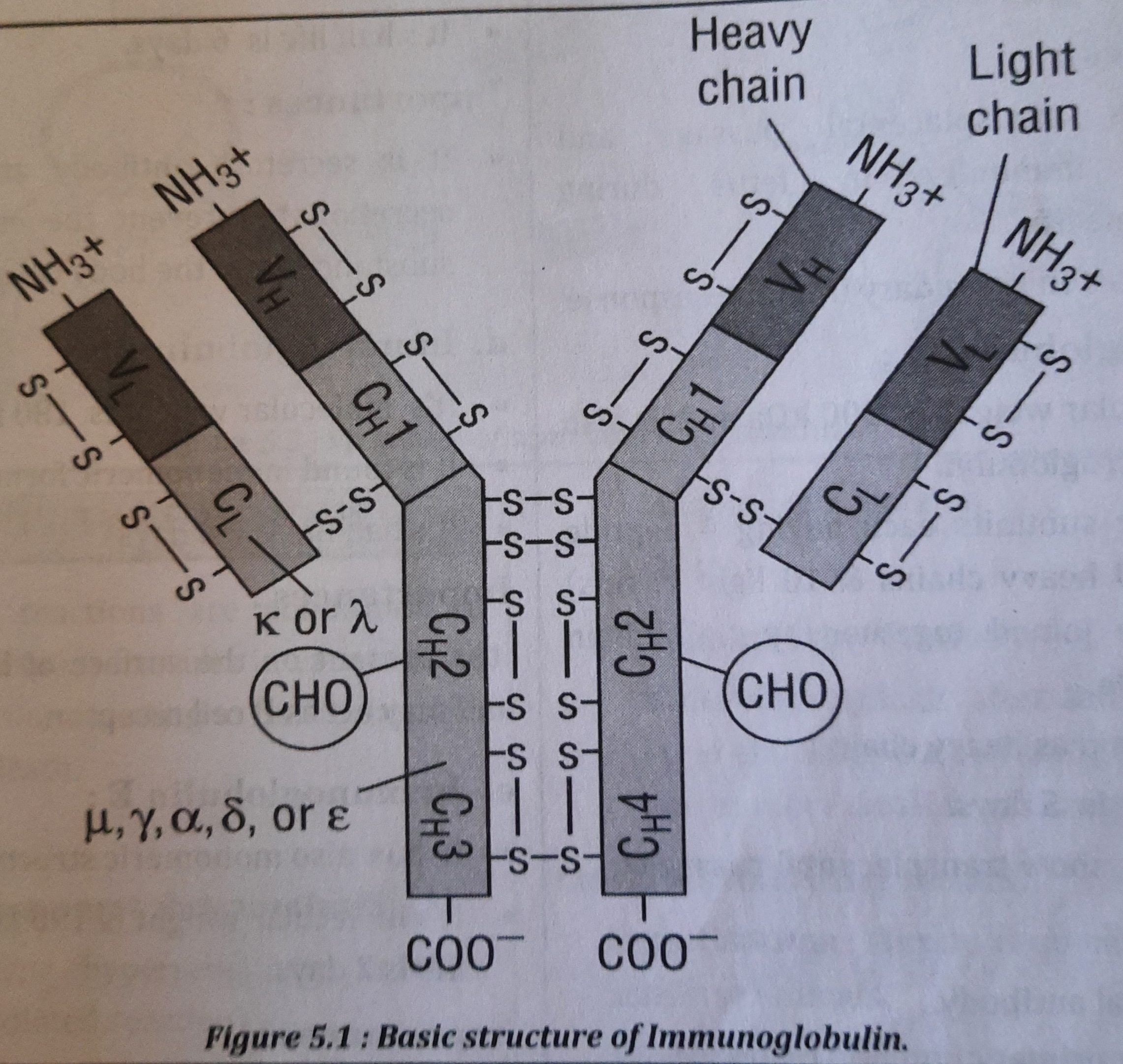
Classification of Immunoglobulin
On the basis of nature of heavy chain, immunoglobulins are classified as below
- Immunoglobulin G(IgG) : contains y-chain
- Immunoglobulin A(IgA) : contains a-chain
- Immunoglobulin M (IgM): contains µ-chain
- Immunoglobulin D (IgD): contains 8-chain
- Immunoglobulin E (IgE): contains ε-chain
a. Immunoglobulin G :
- It constitutes 75-80% of total Ig in circulation.
- It's molecular weight is 150 Kda.
- It has two heavy chains (y-chain) & two light chains.
- It's half life is 23 days
Importances:
- It shows transplacental passage and provides immunity in fetus during intrauterine life.
- It takes part in secondary immune response
b. Immunoglobulin M:
- It's molecular weight is 900 kDa and is also called macroglobulin.
- It has five subunits each having 4 peptide chains (10 heavy chains & 10 light chains) which are joined together by J- chain polypeptide.
- It has µ chain as heavy chain
- It's half life is 5 days.
- It does not show transplacental passage.
Importances:
- It is natural antibody.
- It is seen in primary immune response.
- It is effective for agglutinating more bacteria.
- It is present on the surface of unstimulated B-lymphocytes.
c. Immunoglobulin A:
- It is found in monomer or dimer form. IgA found in secretions is dimer whereas that found in plasma is monomer. J chain connects two monomers to form dimer. It's half life is 6 days.
Importances:
- It is secretory antibody and is found in secretion to prevent the entry of foreign substances into the body cells
d. Immunoglobulin D:
- It's molecular weight is 180 kDa.
- It is found in monomeric form.
- It's half life is 2.8 days
Importances:
- It is present on the surface of B lymphocytes and may act as B cell receptor.
e. Immunoglobulin E:
- It has also monomeric structure.
- It's molecular weight is 190 kDa and it's half life is 2 days.
Importances:
- It mediates allergic reaction.
- It shows high plasma level in helminthic infections.
Hypersensitivity Reactions
Immune responses that are excessive or exaggerated can cause tissue damage, sickness, or even death. These reactions are known as hypersensitivity reactions.
Classification
- On the basis of time onset, they are classified as
- Immediate type hypersensitivity reaction (antibody mediated reaction)
- Delayed type hypersensitivity reaction (T- cell mediated reaction)
- Coombs and gel classified hypersensitivity reactions into:
- Type I hypersensitivity reaction
- Type II hypersensitivity reaction
- Type III hypersensitivity reaction
- Type IV hypersensitivity reaction
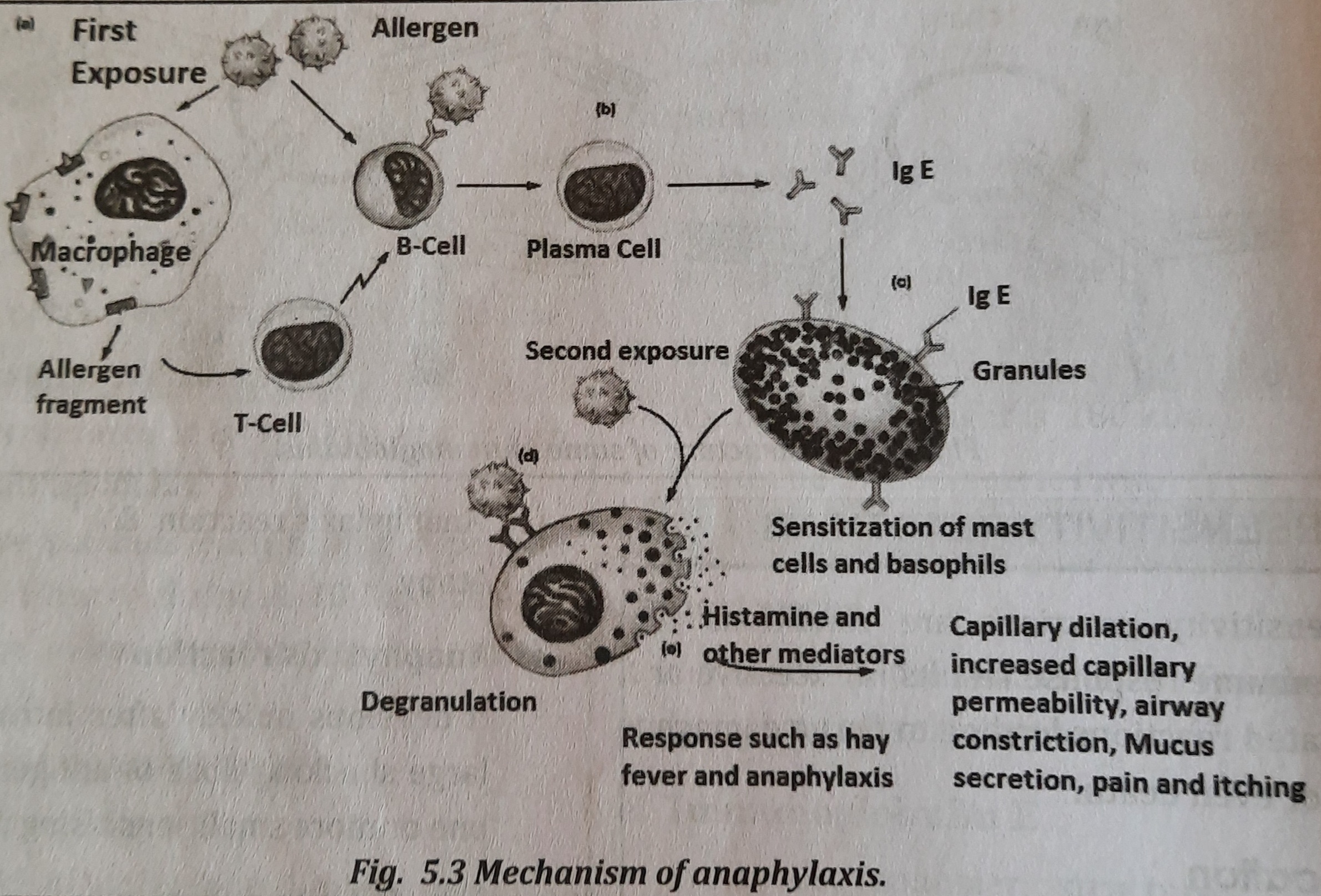
a. Type I Hypersensitivity Reaction:
- It is a type of IgE mediated hypersensitivity reaction.
- It involves two reactions:
- Anaphylaxis reaction &
- Atopy
- Anaphylaxis reaction
- It develops quickly after introduction of large shocking dose of antigen following one or more small sensitising dose.
- It involves following factors:
- Sensitisation: Minute dose of antigen can sensitise animals.
- Waiting period: An interval of 2-3 weeks is required between sensitising and shocking dose.
- Shocking dose: When same antigen is administered, clinical manifestation of anaphylaxis appears.
- Mechanism of anaphylaxis:
- IgE is formed in response to antigenic stimulus and when large amounts are formed, the stage is set for the reaction. Mast cells are normally present in large number in submucosal layer of respiratory tract, gastrointestinal tract, skin and vascular endothelium. IgE molecules are bound to mast cells of tissues and basophils of blood. Fc portion of IgE is bound to mast cells and basophils.
- On subsequent exposure to a large dose of the same antigen, antigen combines with Fab portion of IgE bound to mast cells and basophils. This binding of antigen leads to degranulation of mast cells and circulating basophils and release vasoactive amines that cause anaphylactic reaction.
- Atopy
- It occurs spontaneously in response to substances encountered in the environment in everyday life such as grasses, trees, foods, etc.
- Atopens combine with cell bound IgE molecules which are fixed on the surface of mast cells & basophils in tissues and Ag-Ab complex stimulates the release of mediators that set of the symptoms of atopy.
- Features of Atopy:
- Shows familial distribution
- Reaction occurs at the site of entry of antigen
- IgE mediated reaction
b. Type II hypersensitivity reaction
It is carried out by antibodies that are focused on an antigen found on the cell surface. Through the Fab region, the antibody binds to the antigen. The Fc portion of the antibody is where the complement binds, causing the lysis of cells. So, the lysis of the cells is caused by complement. autoimmune hemolytic anemia, for instance.
c. Type III hypersensitivity reaction:
In tissues, antigen and antibody bind to create the Ag-Ab complex. Polymorphonuclear leucocyte infiltration triggers complement activation, which causes tissue injury.
Two typical type III reactions are:
- Arthus reaction
- The repeated subcutaneous injection of antigen into rabbit causes high levels of antibody in blood. When the same antigen is injected intradermally or subcutaneously in that animal, intense local edema & hemorrhage develop. Same type of reaction appears in man also.
- Serum sickness
- Only a single dose of antigen is sufficient to produce hypersensitivity reaction. The injection of antigen leads to production of antibody. Ag-Ab complex formed may circulate or may be filtered out in important organs & tissues (eg, in kidneys, muscle, lymphnode & joints) leading to damage to the tissue.
d. Type IV hypersensitivity reactions (Delayed type reaction)
It is a slowly evolving reaction (in 24-72 hrs). In type IV, tissue damage events are mediated by T-lymphocytes & not by antibody.
Three type of type IV reactions are:
- Tuberculin type reaction: Tuberculin or purified protein derivative (PPD) is used in this test. Intradermal injection of PPD into sensitised individual causes an erythema, swelling in the skin after 12 hrs.
- Contact dermatitis: Delayed type of hypersensitivity reaction may develop in skin after repeated contact with sensitising materials like drugs, metals(nickel, chromium), simple chemicals (hair dyes, cosmetics, soaps).
- Granulomatous type: It is due to the particles within the macrophages, which the cell unable to destroy.
| Immediate type reaction | Delayed type reaction | |
|---|---|---|
| Timing |
Appears rapidly within minutes |
Appears slowly in 24-72 hours and lasts longer for days |
| Immune response | Antibody mediated | Cell mediated |
| Desensitization | Easy but short lived | Difficult but long lasting |
| Cellular response | Limited polymorphonuclear infiltration | Predominantly mononuclear cell infiltration |
Immunodeficiency disorders
Immunodeficiency disorders are seen when the subject is immunocompromised.
They are classified into two groups:
- Congenital or primary immunodeficiency disorder(PID)
- Acquired or secondary immunodeficiency disorder (SID)
Basic Concept of Immunology in Diagnosis of Viral Diseases
The potent tools of PCR (polymerase chain reaction), flow cytometry, DNA typing, ELISA (enzyme linked immunosorbent assay), and radio-labeled procedures have been made available to immunologists thanks to technological advancements. The immunologist has also long employed precipitation, agglutination, full fixation, and associated processes.
The detection of even the tiniest amount of a biological substance, such as an antigen, is highly suited to immunology. Utilizing substances conjugated with radioactive substances, fluorescent substances, enzymes, and chemiluminescent substances, antibodies made against a particular antigen can also be found.
Immunology has been very useful for the early detection of viral disease, monitor its progression and to assess its prognosis. The antigen-antibody reaction forms the base for the diagnosis of various viral diseases. The immunological techniques are employed for the diagnosis of several viral disease such as Hepatitis, HIV-AIDS.
Things to remember
© 2021 Saralmind. All Rights Reserved.
 Login with google