Oxygen Therapy
Subject: Fundamentals of Nursing

Overview
Oxygen Therapy
Introduction
Oxygen therapy is the administration of oxygen as a medical intervention, which can be for a variety of purposes in both chronuc and acute patient care. Oxygen therapy is the administration of oxygen at concentrations greater than that in room air to treat or prevent hypoxemia. The amount of oxygen the patient uses for inspiration can be increased by providing a supplemental supply via oxygen therapy. Oxygen is a colorless, odorless, tasteless gas which can assist patients in a variety of circumstances and is universally accepted for routine use in hospital setting. It may be classified as an element, a gas, and a drug. O2 is administered whenever there is deficiency in the blood shown by cyanosis. Appropriate levels of oxygen are vital to support cell respiration and cell metabolism. The atmospheric content of oxygen in air is 21%, this amount is adequate for healthy individuals but those certain diseases need an increased oxygen fraction in the gas they breathe. Breathing prescribed oxygen increases the amount of oxygen in the blood, reduces the extra work of the heart, and decreases shortness of breath. The normal amount of O2 in the blood must be in the range of 80 to 100 mm of Hg.
Purposes of Oxygen Therapy
- To increase oxygen saturation in tissues where the saturation levels are too low due to illness or injury.
- To maintain the ability of cells to carry out the normal metabolic function.
- To reduce effects of anoxaemia.
- To decrease respiratory distress.
Factors Affecting Oxygenation
Breathing is dependent on physiological, developmental, environmental and mechanical factors.
- Physiological Factors: Any condition affecting cardiopulmonary functioning direct affects the ability of the body to meet oxygen demands.
- Decreased 02 carrying capacity: Anemia and inhalation of toxic substance decreases the oxygen carrying capacity of the blood by reducing the amount of hemoglobin to transport oxygen.
- Decreased inspired oxygen concentration: With the decline of the concentration of inspired oxygen, the oxygen carrying capacity of the blood decreases e.g. Airway obstruction, High altitude and Hypoventalitation.
- Increased metabolic rate increases oxygen demand e.g. Fever, exercise.
- Hypovolumia: Decreased circulating volume results hypoxia such as dehydration, hemorrhage.
- Alteration of Chest wall motion: Any condition reducing chest wall movement results in decreased ventilation such as Pregnancy, Obesity, Trauma, CNS alterations, Pain, Musculoskeletal Abnormalities, Post-Surgery.
- Developmental Factors: The developmental stage of a client and the normal ageing process affect tissue oxygenation such as Prematurity due to surfactant, Infancy due to recurrent respiratory infection, Older Adult due to atherosclerotic plaques.
Behavioral Factors: Such as Nutrition, Exercise, Smoking, Anxiety/Stress, Substance abuse, Depression.
Environmental Factors: Such as Toxic inhalants, Pollution, Allergens.
Sign and Symptoms of Hypoxia
The degree of hypoxia the patient is experiencing usually determines what signs and symptoms he or she will exhibit. The patient's age, general health, current disease process, and history of chronic illness also play a role in how the patient responds to hypoxia.
In the early stages of hypoxia, the patient is often restless and confused, coughing or wheezing and might report feeling anxious and decrease level of consciousness. The patient's vital signs might also vary from baseline, with heart rate, respiratory rate, and blood pressure elevated. In the late stages of hypoxia, the patient is likely to develop hypotension, bradycardia, and metabolic acidosis. The patient may also develop cyanosis, a bluish discoloration of the skin and mucous membranes.
For The patients who have chronic hypoxia, the manifestations differ. These patients often have clubbing of their fingers and toes, peripheral edema, right-sided heart failure, and oxygen saturation below 87%. It is important to recognize the signs and symptoms of hypoxia and begin oxygen therapy as soon as possible. If hypoxia is left untreated, the patient's condition can deteriorate, resulting in a decrease in activity level, an increase in confusion, a decrease in level of consciousness and possibly coma.
Types of Oxygen Delivery Systems
The types of oxygen delivery systems include:
Compressed Oxygen: Oxygen is stored as a gas in a tank. A flow meter and regulator are attached to the oxygen tank to adjust oxygen flow. Tanks vary in size from very large to smaller portable tanks. This system is generally prescribed when oxygen is not needed constantly (e.g. when it is only needed while performing physical activity).
Liquid Oxygen: Oxygen that is stored in a large stationary tank stays in the home. Liquid oxygen is more highly concentrated, so more oxygen can fit in a smaller tank. A portable tank is available that can be filled from the stationary tank for trips outside the home. Oxygen is liquid at very cold temperature. When warmed, liquid oxygen changes to a gas for delivery to the patient. These tanks are refillable.
Oxygen Concentrators: These devices concentrate oxygen from the air and deliver it to client. An oxygen concentrator is a device that takes oxygen from the room, concentrates it for therapeutic use, and removes other naturally occurring gases. This is not portable and requires electricity to work. A cylinder of oxygen is provided as a backup in the event of a power failure. This system is generally prescribed for The patients who require constant supplemental oxygen or who must use it when sleeping. The benefits of concentrators are that they are less expensive and don't require filling like tanks.
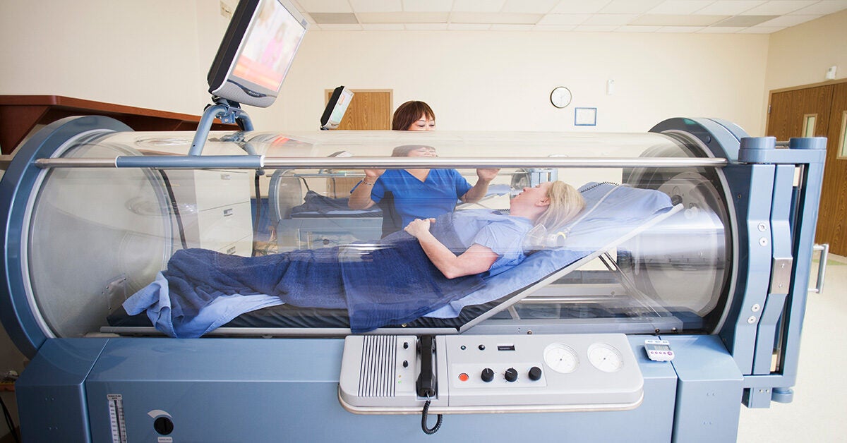
Hyperbaric Oxygen Therapy: Hyperbaric oxygen therapy is the use of high levels of oxygen for treatment of specific diseases. People breathe in pure oxygen in a pressurized room or chamber. In the hyperbaric chambers, the air pressure is increased to three or four times the normal air pressure levels. This increases the amount of oxygen delivered to the body's tissue. This type of oxygen delivery is often used to treat wounds, serious infections, or bubbles of air in your blood vessels.
Sources of Oxygen
Therapeutic oxygen is supplied from a wall outlet or a portable cylinder. A specially designed flow meter is attached to the wall outlet. A valve regulates the oxygen flow in liters per minute. To release oxygen safely and at the desired rate from a cylinder or tank, a regulator is used. The regulator has two gauzes. The one nearest the tank shows the pressure or amount of oxygen in the tank. The other gauge indicates the number of liters per minute of oxygen being released. Oxygen concentrators are another way to provide oxygen. They are used frequently in home situations.
Oxygen Flow Rate
The flow rate of oxygen measured in liters per minute determines the amount of oxygen delivered to the patient. The rate varies depending on the condition of the patient and the route of administration of the oxygen. To regulate the oxygen percentage concentration accurately, samples of the air mixture the patient is actually inhaling should be analyzed every 4 hours. Several types of commercial oxygen analyzers are available. The physician's written order prescribes the rate of oxygen administration. Closely monitoring the flow rate for the patients with chronic lungs conditions is necessary. The chemoreceptors of the patients with chronic lung disease become insensitive to carbon dioxide and respond to hypoxia to stimulate breathing. If excessive oxygen is given, the stimulus to breath is removed; as a result, the patient may stop breathing completely. Arterial blood gas results should be monitored closely for changes. Many times, continuous pulse oximetry also used to monitor the patient receiving oxygen. Oxygen therapy can be delivered using a low flow or high flow system.
Low-flow system includes:
- Simple face mask
- Non re-breather face mask (mask with oxygen reservoir bag and one-way valves which aims to prevent/reduce room air entrainment)
- Nasal prongs
- Tracheostomy mask
High flow systems include:
- Ventilators
- CPAP/BiPAP drivers
- Face mask or tracheostomy mask used in conjunction with an Airvo2 Humidifier
- High Flow Nasal Prong therapy (HFNP)
Humidification
There is no need of humidification with very low flow oxygen (2L/min or less) delivered by nasal cannula; however, as oxygen dries, it dehydrates the respiratory mucous membranes. Humidifying devices are commonly used when oxygen delivered at higher flow rates. The type of humidification device selected will depend on the oxygen delivery system in use and the patient's requirements. Distilled or sterile water is commonly used to humidify oxygen. The humidifier should always be placed at a level below the patient's head so that water from the humidifier does not enter the tubing through which the oxygen is flowing.
Rationale for Humidification
- Cold, dry air increases heat and fluid loss.
- Medical gases, including air and oxygen, have a drying effect on mucous membranes resulting in airway damage.
- Secretions can become thick & difficult to clear or cause airway obstruction.
- In some conditions e.g. asthma, the hyperventilation of dry gases can compound bronchoconstriction.
Oxygen Delivery Devices
A variety of oxygen-delivery devices are available for administering oxygen therapy such as nasal cannula, nasal catheter, transtracheal catheter, simple mask, partial rebreather mask, non rebreather mask, venture mask and tent. Selection of oxygen delivery method depends on age of the patient, oxygen requirements/therapeutic goals, presence of an artificial airway, environment and patient tolerance to selected interface.
Nasal Cannula: A nasal cannula is the device most often used to administer oxygen therapy. It consists of a length of tubing, usually 7 to 14 feet long, with two small prongs to insert into one of the patient's nares. It also has a plastic piece at the neck that slides up under the patient's chin to tighten the tubing and keep it in place. It is available in a range of sizes and can be used for various age groups. A nasal cannula delivers oxygen concentrations of 22% to 50% with flow rates from 1 to 6 L/min through the cannula. The exact concentration inspired depends on the flow rate and on the patient's rate, pattern and the depth of respirations. A nasal cannula is usually used for The patients who are noncritical with minor breathing problems and for The patients who cannot or will not wear an oxygen mask. Because this device administers low- flow oxygen, humidification is rarely required.
Simple Mask: A simple face mask is usually used for the patients who require a moderate flow rate for a short period of time. It is composed of a plastic mask that fits snugly over the patient's mouth and nose. The mask has holes on each side that are used for exhalation and for air entrainment if the flow rate is too low. An adjustable elastic strap that fits over the patient's head holds the mask in place. A piece of tubing connects the mask to the oxygen source. Extension tubing is usually added to allow the patient more freedom of movement. A simple mask has the ability to deliver oxygen concentrations of 40% to 60% with flow rates from 6 to 10 L/min. Because carbon dioxide can build up in the mask at low flow rates, do not use a flow rate lower than 6 L/min with this type of mask. When using this mask, consider humidification to keep the patients' mucous membranes from becoming dry.
Partial Rebreather Mask: It is similar to a simple face mask but is equipped with a reservoir bag for the collection of the first part of the patient's exhaled air. The remaining exhaled air exits through vents. The air in the reservoir is mixed with 100% oxygen for the next inhalation. Thus the patient rebreathes about one third of the expired air from the reservoir bag. This type of mask permits the concentration of oxygen. It delivers oxygen concentrations of 70% to 90% with flow rates from 6 to 15 L/min through the cannula.
Nonrebreather Mask: This device is used to deliver high flow rates and high concentrations of oxygen. Like a simple mask, the nonrebreather mask fits snugly over the patient's mouth and nose. An adjustable elastic strap that fits over the patient's head holds the mask in place. A nonrebreather mask has ports on each side that have one-way valves that keep the patient from breathing in room air to ensure that a high concentration of oxygen is delivered. The mask also has a reservoir bag that is inflated with pure oxygen. Between the mask and the bag is another one-way valve that allows the patient to breathe in the oxygen supplied by the source as well as oxygen from the reservoir. This provides the patient with an oxygen concentration of nearly 100%. A piece of tubing, usually connected to extension tubing, connects the mask to the oxygen source. A nonrebreather mask can deliver oxygen concentrations of 60% to 95% with flow rates from 10 to 15 L/min. When using a nonrebreather mask, do not allow the reservoir bag to deflate. If it does deflate, the patient is likely to breathe in a large amount of exhaled carbon dioxide.
Venturi Mask: A Venturi mask is most often used for critically ill patients who require administration of a specific concentration of oxygen. It consists of a mask with holes on each side that allow exhaled air to escape. At the base of the mask are color-coded entrainment ports that are adjustable to allow regulation of the concentration of oxygen administered. A Venturi mask can deliver oxygen concentrations from 24% to 60% with flow rates from 4 to 10 L/min. Because this device delivers a precise oxygen concentration and carbon dioxide buildup is minimal, it is commonly used for The patients who have COPD. Humidification is usually unnecessary with this device.
Face Tent: A face tent is often used as an alternative to an aerosol mask, especially for the patients who report feeling claustrophobic with an aerosol mask. It provides oxygen to nose and mouth without the discomfort of mask. It is composed of a soft mask that fits under the patient's chin and loosely covers the mouth and nose. An adjustable elastic strap holds it in place. A face tent delivers oxygen concentrations of 28% to 100% with flow rates from 8 to 12 L/min. This device is convenient for delivering both humidification and oxygen; however, it is difficult to control the concentration of oxygen administered since the actual concentration of oxygen depends on the rate and depth of the patient's respirations.
Oxygen Tent: Oxygen tents and hoods are usually used for pediatric The patients who have airway inflammation, croup, or other respiratory infections. The oxygen tent consists of a canopy that surrounds the child. It provides oxygen, humidification, and a cool environment to help control body temperature. The oxygen hood consists of a disposable vinyl box that fits over the child's head. It provides warm humidified oxygen at a specific temperature. When using a hood, it is important to ensure that there is enough space between the curve of the hood and the child's neck to allow carbon dioxide to escape. An oxygen hood delivers a 28% to 85% oxygen concentration varying with the flow rate, which can be set at 5 to 12 L/min. An oxygen tent can provide oxygen concentrations of up to 50% with flow rates from 10 to 15 L/min.
Manual Resuscitation: A manual resuscitation bag is used to provide high concentrations of oxygen to a patient prior to a procedure, such as suctioning or intubating, and during respiratory or cardiac arrest. It can also be used to assist The patients who are breathing but not adequately. It consists of a mask, a self-inflating bag that is compressed to ventilate the patient, and an oxygen port where the oxygen tubing is connected. It might also have an adapter that fits onto an oxygen port where the oxygen tubing is connected. It might also have an adapter that fits onto an endotracheal tube when it is going to be used for an intubated patient. The mask fits over the patient's nose and mouth and has a soft air-filled cushion around the mask that forms an airtight seal when placed on the patient's face. The apex, or narrow portion of the mask, is placed over the nose and the base, or broader portion of the mask, over the mouth.
Self-inflating Bag: The self-inflating bag is made of a firm, rubber-like material that is manually compressed to give the patient "a breath." The bag has an oxygen port where oxygen tubing can be connected if the patient requires high concentrations of oxygen. There is also a valve on the bag that ensures one-way pressure into the mask and then allows the bag to reinflate from ambient air or from an oxygen source.
Tracheostomy Mask: A tracheostomy mask sometimes refers to as a tracheostomy collar, is a small mask that fits over the patient's tracheostomy site. An adjustable elastic strap that fits around the patient's neck holds it in place. The mask has an exhalation port that remains patent at all times and a port that connects to the oxygen source with large- bore tubing. The flow rate is usually set at 10 L/min, with a nebulizer set at the appropriate oxygen concentration. The patients who have artificial airways require continuous humidification since the airway bypasses the normal filtering and humidification process of the nose and mouth. The two devices most commonly used are a t-tube, also called a Briggs adaptor, and a tracheostomy mask. A t-tube is a t-shaped device with a piece that connects the oxygen source to the artificial airway (endotracheal tube or tracheostomy). The recommended flow rate when using a t-tube is 10 L/min, with a nebulizer set at the appropriate oxygen concentration.
Noninvasive Ventilation: An alternative to mechanical ventilation, it is used to maintain positive airway pressure and to improve alveolar ventilation without the need for an artificial airway. It is commonly used for the patients who have congestive heart failure, sleep disorders, and pulmonary diseases to improve oxygenation, reduce and reverse atelectasis, reduce pulmonary edema, and improve cardiac function. The two types of noninvasive ventilation are CPAP and Bi PAP. Continuous positive pressure ventilation (CPAP): It provides a set positive airway pressure throughout the patient's breathing cycle. It is commonly used for the patients who experience sleep apnea because the continuous positive pressure keeps the airway open and prevents the upper airway from collapsing. The usual CPAP pressure is between 5 and 20 cm of water.
Bi level positive airway pressure (Bi PAP): It provides assistance during inspiration and keeps the airway from closing during expiration. The benefits of BiPAP include an increase in the amount of air in the lungs at the end of expiration, reduced airway closure, and improved oxygenation.
Indication for O₂ Inhalation
Administration of oxygen is based on the provider's assessment of the patient. Oxygen therapy is indicated for the patients who are at risk for developing hypoxia. Oxygen may be used in following conditions
- Respiratory problems: Obstructed air way due to growth, enlarged thyroid asthma, pneumonia, chest trauma, pulmonary edema.
- Cardiac problems: Cardiac failure, cardiac insuffiency, anaemia.
- Hypervolemia, excessive fluid loss, after severe haemorrhage.
- Shock
- Increased metabolic rate during fever, exercise
- Seizure patients
- Patient's under anesthesia and Post-operative period.
- Altered Mental Status
- Asphyxia.
- Poisoning with chemical that alter the tissues ability to utilize O2 e.g. carbon monoxide poisoning.
Articles
- O2 cylinder with flow meter connected to O₂ cylinder and regulator, humidifier with sterile distilled water.
- Cylinder stand.
- Key.
- Nasal cannula, O2 mask with connecting tubes.
- Gauze pads/cotton.
- A bowl with plain water to check O2 flow from tube and to make it soft.
- Tape and scissors if necessary.
Method of O₂ Therapy
| S.N. | Nursing Action | Rationale |
| 1 | Determine need for oxygen therapy in the patient and verify the order for the therapy. | Reduces the risk of error. |
| 2 | Perform an assessment of vital signs, level of oxygen, lab values, etc. and record. | Provides a baseline data. |
| 3 | Place the patient in comfortable position. Assist the patient on semi-fowler's position if possible. | Semi-fowler's position permits easier chest expansion and breathing. |
| 4 | Explain procedure to the patient and inform them how to cooperate. | Reduces anxiety and enhance cooperation. |
| 5 | Wash hands. | Reduces risk of transmission of microorganisms. |
| 6 |
Arrange the required articles. Set up O₂ equipment and humidifier. Fill the humidifier up to the mark on it. Attached flow meter to source. Attached humidifier bottle to base of the flow meter. Attach tubing and nasal cannula/face mask to humidifier. Check the condition of O2 pipe and flow meter. Regulate flow meter to prescribed level. Ensure proper functioning by checking for bubbles in humidifier or feeling O2 at the outlet.
|
Filling humidifier above this level will cause water to enter into tubing. Flow meter helps in monitoring and regulating oxygen flow. Humidification helps to prevent drying of nasal mucosa. |
| 7 | Clean the nostril with swab stick, if the nostrils are blocked with secretion. | Ensures correct administration. |
| 8 |
To administer O2 by nasal cannula Check nasal cannula by dipping in a bowl of water and note the O2 coming out of cannula. Place tips of cannula to the patient's nares and adjust straps around ear for sung fit. |
Ensures the flow of oxygen. |
| 9 |
To administer O2 by face mask Guide the mask to the patient face and apply it from nose down ward. Fit the metal piece of mask to confirm to the shape of nose. |
The mask should mould to face so that very little oxygen escapes into eyes or around cheeks. |
| 10 | Secure elastic band around the patient's head. | Ensures comfort of patient. |
| 11 | Apply padding behind ears as well as scalp where elastic band passes and inspect skin behind the ear periodically. | Padding prevents skin irritation/breakdown |
| 12 | Ensure that safety precautions are followed. | Reduces the risk of error. |
| 13 |
Inspect the patient and equipment frequently for flow rate, clinical condition, level of water in humidifier, etc. Remove the mask and dry the skin every 2-3 hours of oxygen administered continuously. Remove and clean the cannula with soap and water, dry and replace every 8 hours. Assess nares at least every 8 hours. |
Identifies complications if develop.Presence of cannula causes irritation and dryness of the mucous membrane. |
| 14 |
Administering O2 using O2 tent Select the smallest tent and canopy. Tuck the edge of the tent under the mattress securely. Pad the metal frame that supports the canopy. Flush the tent with O2 after it has been opened for a period of time to increase the concentration of the gas, and then reset the flow meter to the original level. Analyze and record the tent atmosphere every 1-2 hours. Maintain a tight fitting canopy whenever possible; provide nursing care through the sleeves or pockets of the tent. Check the child's temperature routinely. Moisture accumulation may result in hypothermia.
|
That will achieve the desired concentration of O2 and maintain the patient's comfort. This is especially important if the child restless and can dislodge the tent by pulling the covers loose which leads to oxygen leakage. Padding prevents skin irritation and injury. Concentrations of 30% to 50% can be achieved in well maintained tents. Moisture accumulation may result in hypothermia. |
| 15 | Wash hands. | Reduces the risk of transmission of microorganisms. |
| 16 | 'No Smoking' sign should be pasted in the unit. | Oxygen helps in combustion. |
| 17 | Documents time, flow rate and observations made on the patient. | Serves as a communication between staff members. |
Patient Assessment and Documentation
- Assess the airway and optimize airway position (e.g. head tilt, chin lift) as necessary.
- Clinical assessment and documentation including but not limited to: cardiovascular, respiratory and neurological systems should be done at the commencement of each shift and with any change in patient condition.
- Check and document oxygen equipment set up at the commencement of each shift and with any change in patient condition.
- Hourly checks should be made for the following:
- oxygen flow rate
- patency of tubing
- humidifier settings
- Hourly checks should be made and recorded on the patient observation chart for the following (unless otherwise directed by the treating medical team):
- heart /respiratory rate
- respiratory distress (descriptive assessment i.e. use of accessory muscles/nasal flaring oxygen saturation)
- Continuous pulse oximetry is recommended for the patients who are severely ill, and who are likely to have rapid and clinically significant drop in oxygen saturations when the oxygen therapy is disconnected.
- The venturi mask will have color coded inserts that list the flow rate necessary to obtain the desired percentage of oxygen.
Flow rates and oxygen concentrations delivered using venture mask
| Nozzle color code | Flow rate L/minute | Concentration of oxygen to be delivered |
| Blue | 3Lpm | 24% |
| Yellow | 6Lpm | 28% |
| White | 8Lpm | 31% |
| Green | 12Lpm | 35% |
| Pink | 15Lpm | 40% |
| Orange | 15Lpm | 50% |
Precautions for Oxygen Therapy
- O₂ must be administered with care.
- Since O2 acts as a drug, it must be prescribed and administered in specific dose in order to avoid O2 toxicity. The dosage of O2 is started in terms of concentration and rate of flow
- Change the nasal catheters at least 8 hours or more often.
- When O₂ therapy is discontinued, it should be done gradually.
- Watch the patients receiving O₂ therapy continuously to defect the early signs of O toxicity.
- Promote safety measures; inform that smoking is not permitted in the area of O2 use. Place "NO SMOKING" signs in patient's room or door.
- Avoid open flames in the patient's room.
- Place more fire alarms throughout the home to help prevent serious complications.
- O2 cylinder should be stored at a low temperature because high temperature can cause expansion of the gas with consequent loss of gas through safety valve.
- Never increase or decrease the flow of O2 when the nasal cannula is in the patient's nostrils.
- Do not use electrical appliances close to O2.
- Ensure the humidifier bottle is always at least 1/3 full with sterile water.
- Oil or grease should not be used on the regulator and cylinder because in the presence of high concentration oil is likely to catch fire and the cylinder may explode.
- Only store oxygen in an area where air moves freely around the tank. Do not store it in a trunk or a small closet.
- Do not use drugs or alcohol while taking oxygen therapy, as they can slow your breathing.
- Naver cover the oxygen therapy device with any material.
- Turn off oxygen immediately when not in use. Oxygen is heavier than air and will pool in fabric making the material more flammable. Therefore, never leave the nasal prongs or mask under or on bed coverings or cushions whilst the oxygen is being supplied.
Potential Complications of Oxygen Therapy
Oxygen is not addictive and causes no side effects when used as prescribed. Complications from oxygen therapy used in appropriate situations are infrequent.
- Respiratory depression/CO₂ Narcosis: This occurs in the patients who have chronic respiratory obstruction or respiratory insufficiency which results in hypercapnea (i.e. raised PaCO2). In these patients the respiratory centre relies on hypoxaemia to maintain adequate ventilation. If these patients are given oxygen, this can reduce their respiratory drive, causing respiratory depression and a further rise in PaCO2.
- Pulmonary Atelectasis: absorption atelectasis is the most serious complications of oxygen overuse.
- Pulmonary Oxygen Toxicity: High concentrations of oxygen (>60%) may damage the alveolar membrane when inhaled for more than 48 hours resulting in pathological lung changes.
- Sub Sternal Pain: characterized by difficulty in breathing and pain within the chest, occurring when breathing elevated pressures of oxygen for extended periods.
- Oxygen Delivery equipment may present other problems. Perforation of the nasal septum as a result of using a nasal cannula and non-humidified oxygen has been reported.
- In addition, bacterial contamination of nebulizer and humidification systems can occur, possibly leading to the spread of pneumonia
Things to remember title
© 2021 Saralmind. All Rights Reserved.
 Login with google