Human Eye
Subject: Science

Overview
The human eye is an optical device that creates images of objects by refracting light via a convex lens. It consists of the cornea, pupil, eye lens, ciliary muscles, retina, and optic nerve. Accommodation of the eye involves adjusting the lens's thickness based on the object's distance. Shortsightedness or myopia, causes blurry images of far-off objects due to the lens focusing in front of the retina. Long-sightedness or hypermetropia, results in light from surrounding objects concentrated behind the retina, making nearby objects appear hazy but producing a crisp image of distant objects. Contact lenses are another option for repairing vision defects. Cone cells in the retina, which are in charge of detecting colors, have abnormalities that lead to color blindness. Night blindness occurs when the rod cells in the retina malfunction, making objects in low light invisible. Corneal injuries, such as scratches, wounds, and infections, can lead to corneal disorders and vision issues. Corneal transplantation is a treatment for corneal degeneration, which is the second most common cause of blindness in the country.
The human eye is an optical device that naturally creates images of objects by refracting light via a convex lens. The spherical shape of the eye is attributed to the aqueous humor and vitreous humor within it, as depicted in the picture. The following describes some of the key components of the human eye.
- Corneal
The transparent layer in front of the eye that lets light in is called the cornea. It facilitates light focus on the retina. The cornea bends (refracts) light more than any other portion of the eye when light rays from objects travel through the air, cornea, aqueous humor, lens, and vitreous humor before arriving at the retina. The cornea functions as the eye's primary lens as a result.
- Pupil
The black aperture in the center of the eye where light enters the eye is called the pupil. Its dimensions vary depending on how brilliant the light is. The iris, or outer layer of muscles surrounding the pupil, is what determines the pupil's size. The iris muscles contract and the pupil widens in low light, while the iris muscles relax and the pupil shrinks in bright light. As a result, even in areas with very little or no light, regulated light can enter the eye.
- Eye lens
The translucent convex lens of the human eye is composed of a naturally occurring protein known as crystalline. In order to converge onto the retina, the light rays that are refracted from the cornea are further refracted in the lens.
- Ciliary Muscles
The flexible muscles that alter the thickness of the eye lens are called ciliary muscles. Convergence of light beams onto the retina is maintained by the ciliary muscles that are linked to the lens. The focal length and curvature of the lens alter as the ciliary muscles contract and relax.
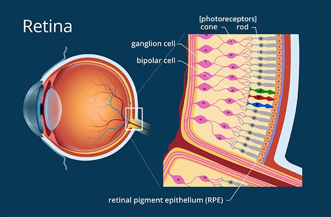
- Retina
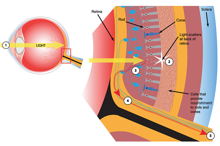
The layer of light-sensitive cells in the back of the eye, called the retina is responsible for detecting light's color and brightness. An image of the object is created when light beams are directed onto the retina. Rod cells and cone cells are the two cell types found in the retina. Rod cells transform light into electrical signals and aid in determining light brightness, whilst cone cells aid in the detection and identification of colors.
- Optic Nerve
The optic nerve is a collection of nerve cells in the retina that sends electrical signals to the brain carrying information about the image.
The convex lens on the retina creates an inverted image, but our brain flips it so that we view the things correctly. The creation of an image is feasible due to the intricate structure of the human eye and the roles played by its various components, as previously described. Whether the light is coming from a nearby or distant object, it must constantly be refracted and precisely focused on the retina to generate a distinct image on the retina.
Accommodation of Eye
When we gaze at the letters in a book we are holding, the object distance gets shorter; when we look at the clouds in the sky, the object distance gets longer. The image distance, or the distance between the lens and the retina, doesn't change since the retina forms the images of things at different distances. In this case, the lens's curvature must alter to cause the light rays to roughly converge in order to provide a clear image. The eye's cillary muscle can adjust the lens's thickness to compensate for this.
Looking at an item in the distance causes the cillary muscles to relax. In this case, the focal length grows and the lens becomes thinner, or its curvature reduces. We are able to see the far-off objects clearly because the incoming light is focused on the retina. In the same way, the ciliary muscles contract when one looks at a nearby object. Under this circumstance, the focal length shortens, the lens thickens, and its curvature increases. Consequently, the inbound light beams from the adjacent object undergo more refractive effects and concentrate on the retina. Accommodation of the eye refers to the process of modifying the focal length of the eye lens based on the distance of the object from the eye through ciliary muscle contraction and relaxation.

Far Point and Near Point of Human Eye
The farthest distance at which an object may be clearly seen from the eye is known as the far point. This point is located at infinity for a typical eye. The eye's lens narrowest and longest focal length occurs when it is focused on a far-off object. Similarly, the ciliary muscles contract to their greatest extent when the item is moved to a specific distance from the eye. The ciliary muscles cannot contract any more if the item is moved closer, making it impossible for the retina to display a crisp image.
The near point of the eye is the closest distance at which it is able to view objects well. The near is 25 centimeters away from the eye in a typical eye. The least distance of distinct eyesight is another name for this distance. The eye lens has the largest curvature, or it becomes the thickest and has the shortest focal length, while staring at an object point at the closest point.
Depending on the individual's age and eye health, the near point of the eye might change. The range of human vision for an individual without any visual impairment is 25 cm to infinity (∞). The near and distant points of the eyes are different for people with visual impairments than they are for people with normal vision.
Defects of Vision
A book must be moved closer or farther away from the eyes until the letters are visible if they appear fuzzy while held at a regular distance. As seen in figure 10.66, an object's light rays must focus on the retina to generate a crisp image in order to be seen clearly, whether it is close by or far away. If not, the object will seem hazy. When things appear blurry to humans that is, when distinct images of these items cannot be formed on the retina this occurrence is referred to as a deficit of vision.
Types of Defects of Vision
- Shortsightedness or Myopia
Images of far-off things appear fuzzy to people with shortsightedness because the light rays from those items are focused in front of the retina. Nonetheless, the retina produces a sharp vision for objects that are closer. Shortsightedness is the condition where a person can clearly see objects close to them but not those far away. There are two main causes of this issue:
(a) In certain individuals, the eyeball enlarges and the distance between the retina and lens grows. The parallel rays from far-off objects are therefore directed in front of the retina.
(b) The lens cannot be narrow enough to allow for distant objects to be seen if the ciliary muscles do not contract sufficiently. As a result, the lens's focal length is reduced below what is necessary, focusing the parallel light rays from far-off objects in front of the retina. Myopia can occasionally be caused by both the cornea's and the lens's curvatures. The far point for myopic eyes is not at infinity, but rather a certain distance from the eye.

Correction of Shortsightedness
If the parallel rays arrive in the eyes somewhat before they enter, the shortsightedness issue in that eye may be resolved. As seen in figure 10.67, a diverging appropriate focal length must be maintained in front of the eye for this. As a result, that lens diverges the incoming parallel light beams from the far-off object before they reach the eye. These divergent rays are refracted via the cornea and lens of the eye, focusing on the retina where the object appears clear.

In order to precisely focus rays diverged by the lens on the retina and make distant objects appear to be coming from the far point F of the deficient eye, the focal length of the lens used in the spectacles to remedy the defect is chosen.
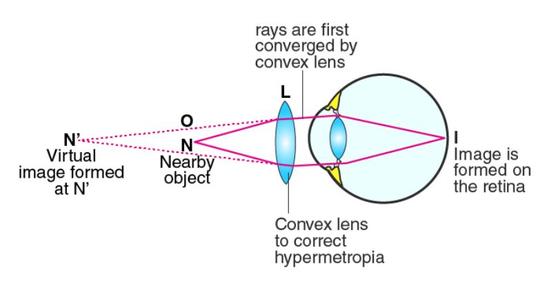
- Long-sightedness or Hypermetropia
When an individual has longsightedness, light from surrounding things is concentrated behind the retina, making nearby objects appear hazy. However, the retina forms a crisp image of the distant object. Long-sightedness is the condition in which one can see adjacent items well while distant objects appear hazy. There are two main causes of this issue:
(a) The distance between the lens and the retina reduces in certain people because their eyeballs become overly round. As a result, light beams from surrounding objects focus behind th
e retina.
(b) The lens cannot be made thick enough if the ciliary muscles in the eye are unable to contract sufficiently while the eye is focused on adjacent objects. As a result, the focal length increases and light beams from surrounding objects focus behind the retina. For people who are long-sighted, the close point is farther away from the eye than 25 cm, as Figure 10.68c illustrates. This issue typically manifests in advanced age.

Correction of Longsightedness
The issue for longsighted eyes can be resolved if the light beams from the surrounding object converge to some In order to do this, a converging lens, also known as a convex lens, should be positioned in front of the eye so that light beams from surrounding objects are somewhat converged by the lens before entering the eye. The surrounding things become sharply visible when those rays focus on the retina after passing through the cornea and the eye lens. The convex lens used in the spectacles to correct longsightedness has a focal length that is chosen so that when light converges on the retina, it appears as though it is coming from the near point N of the longsighted eye.
Other Ways for Correction of Defect of Vision Apart from Using Spectacles
(a) Contact Lens
A contact lens is a tiny, synthetic lens that is applied to the cornea's surface to repair visual impairments. It covers the iris and pupil of the eye and rests above the thin layer of tears on the cornea. This causes the light beams to refract just enough before they hit the eye lens. When performing sports or in other situations where wearing glasses is not desired, contact lenses with powerless sunglasses are a beneficial option.
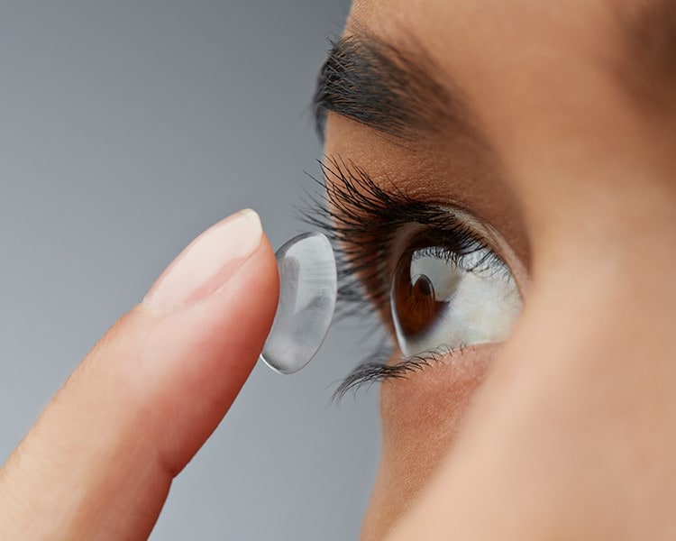
Both wearing glasses and wearing contact lenses has perks and cons. Using glasses is quite simple. It is equipped with premium lenses that come in various sizes. High-power lensed spectacles are bulky, though, and glancing right and left through them distorts peripheral vision. However, since contact lenses move with the cornea, this issue does not arise with them. Low power contact lenses can be used in place of thick glass lenses since they are much closer to the eyes.
Compared to glasses, contact lenses need to be handled with more care. There are precise methods that they must be cleaned and stored. Before being used again, contact lenses need to be cleaned and sterilised. Always wash your hands before putting on and taking out your contact lenses to avoid infection. Wearing contact lenses to bed is not recommended. The cornea may be impacted by incorrect contact lens use.
The cornea of a myopic person's eye is seen in Figure getting somewhat flattened by focussing a laser beam on its center. As a result, the myopia issue is resolved and light beams passing through the cornea are slightly less refracted than they were previously. On the other hand, longsightedness can be resolved if the laser beam is directed toward the cornea's margins, which raises the central portion of the cornea somewhat. In this sense, laser eye surgery refers to the procedure of treating vision impairments by reshaping the cornea using a particular kind of ultraviolet laser, known as an excimer laser.phone
The outermost layer of the cornea is the target of laser eye surgery. A variety of laser eye surgery treatments exist, the most often used being LASIK (Laser-assisted in situ keratomileusis). Here, a flap is removed by the laser beam that is directed toward the cornea. The cornea is reshaped by flipping the cutting flap.
Other Problems Related to the Eye (Apart from Defects of Vision)
(a) Cataract
 People have foggy lenses as a result of the crystalline proteins in their lenses sticking together as they age. As a result, the objects appear hazy because the light from them does not adequately reach the retina. The clear quality of the lens is lost as its opacity rises, leading to eventual total blindness. A cataract is a gray area that develops in the pupil of the eye as a result of lens cloudiness. Most cases of cataracts occur in older adults.
Some people develop spectacles before they reach old age because to factors like excessive blood sugar, smoking cigarettes, and UV radiation from the sun. Surgery is used to treat cataracts.
An incision is made along the cornea's border in order to perform cataract surgery. By passing an ultrasound through the hole, the hazy lens is broken up into small fragments, which are then removed with the use of a suction tube. An acrylic or silicone intraocular lens is retained in its stead.
In 1995, Nepali ophthalmologist Dr. Sanduk Ruit created an incredibly affordable intraocular lens that revolutionized cataract therapy.

(b) Colour Blindness
The color we experience is determined by the color-identifying cells in the retina as well as the color light waves of that color. The figure illustrates how the blue cells in the retina of the eye absorb the blue light wave reflected from an object's surface when one is staring at anything blue. The observer's brain then receives the notification that the color blue has been noticed. There are millions of these three different kinds of cone cells in the retina: blue, red, and green cone cells. Colors are divided by these cells. We lose the ability to identify colors if the retina's cells stop functioning. Red and green hues cannot be distinguished by those who have red-green color blindness. Therefore, color blindness is the incapacity of the eyes to discriminate between colors as a result of abnormalities in the retina's cone cells. Hereditary factors are the main cause of this. In addition, mutations and damaging radiation can kill cone cells, which results in color blindness.
(c) Night Blindness
We can see objects in low light thanks to the rod cells in our retina. For instance, one may still make out the arrangement of the furniture in a room even in extremely low light. Sometimes, the rod cells malfunction and things in extremely dark environments are invisible because of illness, injury, inadequate nutrition, or a host of other factors. The inability to see well at night or in dimly lit areas is known as night blindness, or nyctalopia. This represents the retina's issue. Since a form of protein and vitamin A are the building blocks of the rhodopsin pigment found in the rod cells of the retina, a deficiency in vitamin A causes deficiencies in this pigment. Thus, one of the primary reasons of night blindness is a deficiency of vitamin A in the body. Hereditary factors may also contribute to this illness.
Effects of Injuries to the Cornea in the Eyes
Light in the eye is refracted in part by the cornea. The cornea is where the greatest refraction occurs. The cornea has a refractive index of 1.376 and a converging power of roughly +43 D. It makes up over two thirds of the eye's total capacity for light refraction. Therefore, it is necessary to shield the cornea of the eye from scratches, wounds, and injuries. Occasionally, rubbing the eyes with palms repeatedly can cause the cornea to become scratched when sand or dust gets into the eyes during a storm. In a similar vein, the cornea can be injured by stones, metal fragments, accidents, etc. Corneal infections can result from wearing contact lenses for extended periods of time or from not cleaning them properly. Corneal infections brought on by bacteria, viruses, or fungi can result in corneal disorders and vision issues. Keratitis, or corneal ulcer, is brought on by a corneal infection. People will become blind if they do not receive prompt treatment for their corneal ulcer. Visual impairment is also brought on by fluid buildup between the layers of the cornea, which is known as corneal edema. Similar to this, keratoconus, which happens when the cornea's surface takes on a conical form, first results in shortsightedness before eventually causing the eye to lose its ability to see. Treatment for normal corneal issues can be curative. If therapy is unable to restore the cornea, it may need to be replaced.
Corneal Transplantation
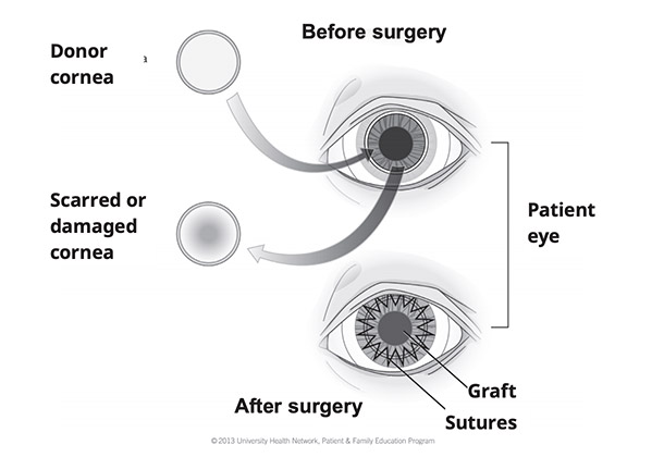
Corneal degeneration is the second most common cause of blindness in our nation, after cataracts.
People who were born with puffed eyes and those whose corneas were harmed by various illnesses, traumas, etc. but did not receive prompt medical attention can both benefit from corneal replacement. Cornea transplantation is the technique by which a healthy cornea is surgically replaced with a damaged one, as illustrated in Figure.
In order to allow others to use their cornea after they pass away, some people donate their eyes to an eye bank. Following the passing of eye donors, medical professionals take only the cornea and securely store it. Within eight to twelve hours of the donor's death, the cornea is extracted from the eye and the eyelids are correctly closed to shield it from the sun. The quality of the cornea improves with prompt removal following death. To collect, store, and distribute corneas, Tilganga Eye Institute in our nation founded the Nepal Eye Bank.
Things to remember title
- The human eye is an optical device that creates images of objects by refracting light via a convex lens. It consists of the cornea, pupil, eye lens, ciliary muscles, retina, and optic nerve.
- Defects of vision can cause blurry images or deficits of vision.
- Shortsightedness or myopia, causes blurry images of far-off objects due to the lens focusing in front of the retina.
- Correction of shortsightedness involves maintaining a diverging focal length to focus light beams before reaching the eye.
- Long-sightedness or hypermetropia, results in light from surrounding objects concentrated behind the retina, making nearby objects appear hazy but producing a crisp image of distant objects.
- Longsightedness can be corrected by using a converging lens to converge light beams from surrounding objects.
- Contact lenses are another option for repairing vision defects.
- Other eye problems include cataracts, which cause foggy lenses and blindness. Surgery is used to treat cataracts, which can be treated with an incision along the cornea's border.
- Cone cells in the retina, which are in charge of detecting colors, have abnormalities that lead to color blindness.
- Night blindness occurs when the rod cells in the retina malfunction, making objects in low light invisible.
- Corneal injuries, such as scratches, wounds, and infections, can lead to corneal disorders and vision issues.
- Corneal transplantation is a treatment for corneal degeneration, which is the second most common cause of blindness in the country.
- Corneal degeneration can be treated with cornea transplantation, which involves surgically replacing a healthy cornea with a damaged one.
© 2021 Saralmind. All Rights Reserved.
 Login with google