Heart
Subject: Science

Overview
The primary organ of the circulatory system, the heart, is in charge of delivering blood to all of the body's tissues and cells. It is made up of heart muscle and has the pericardium, a protective membrane, surrounding it. There are four valves in the heart: the bicuspid, mitral, tricuspid, and pulmonic valves. Heart attacks, which cause agonizing agony and necessitate emergency care, are abrupt disturbances in the blood flow to the heart tissues. Numerous tests, such as those that measure blood pressure, temperature, and pulse, can be used to diagnose heart attacks. Open-heart surgery, coronary artery bypass surgery, and coronary angioplasty and stenting are just a few examples of the medical procedures used to open blocked arteries. A stethoscope can measure the heart rate by listening to the sound that the heart muscle makes when it contracts and relaxes regularly.
The circulatory system's main organ is the heart. Every cell and tissue in our body is supplied with blood via it. The heart, which is located between the two lungs in the thoracic cavity, is composed of cardiac muscle. It has a little leftward inclination. The pericardium, a double-layered protective membrane, envelops the heart. The fluid known as pericardial fluid fills the area between these two layers. It shields the heart from shocks and damage from the outside world. The size of the human heart is the size of the owner's closed fist, and it weighs about 300 grams on average.
The cardiac muscles make up the triangular or conical, muscular organ known as the heart. It regularly contracts and expands to circulate blood throughout the body. Blood is pumped through blood vessels and reaches each cell and tissue of the body. The left side of the thoracic cavity contains around two-thirds of the heart. A hair-like, very fine network of capillaries surrounds the heart. The coronary artery supplies oxygenated blood to the heart, while the coronary sinus vein transports deoxygenated blood to the right auricle of the heart.

Internal Structure of the Heart and Blood Circulation
The heart has four chambers in humans. The bottom two chambers are referred to as ventricles, while the top two are known as auricles or atria. A robust, muscular septum that separates the left and right chambers of the heart also prevents oxygenated and deoxygenated blood from combining inside the chamber. The right auricle, right ventricle, left auricle, and left ventricle are the names of the four chambers of the heart.  These chambers are linked to blood arteries, which are used by the heart to pump and receive blood. While the ventricles carry blood to different parts of the body, the auricles receive blood from different sections of the body. The heart exerts tremendous pressure when pumping blood to various parts of the body. The walls of the ventricles are thicker than those of the auricles in order to handle this kind of pressure. The left ventricle exerts tremendous pressure when pumping blood to different organs, while the right ventricle just pumps blood up to the lungs. As a result, the left ventricle's wall is thicker than the right ventricle's wall.
The inferior and superior vena cava are the blood veins that supply the rig auricle of the heart with blood that is either polluted or deoxygenated. Impure blood is brought from the higher regions of the body by the superior vena cava and from the lower regions of the body by the inferior vena cava. Pure blood flows from the left ventricle to the left ventricle and impure blood from the right auricle to the right ventricle when the auricles contract. While the pulmonary vein carries oxygenated blood to the left auricle of the heart, the pulmonary artery carries deoxygenated blood to the lungs for oxygenationThe pulmonary vein is the only vein in the human body that carries oxygenated blood, and the pulmonary artery is the only artery that does so. The aorta is used to pump blood from the left ventricle to different areas of the body once the pure blood has been transferred from the left auricle to the left ventricle.
The heart contains four valves. The tricuspid valve is the valve that is located between the right auricle and the right ventricle of the heart. Blood flows from the right auricle to the right ventricle when this valve is opened. The bicuspid valve, also known as the mitral valve, is the valve that is located between the left auricle and left ventricle of the heart. This valve opens to allow pure blood from the left auricle to enter the left ventricle. When the auricles contract collectively, both valves open simultaneously, and both ventricles contract after the auricles contract. The pulmonic valve, which opens as the right ventricle contracts and closes the tricuspid valve, allows deoxygenated blood to be pushed to the lungs via the pulmonary artery, where it is oxygenated. Similar to this, when the left ventricle contracts, the bicuspid valve closes, the aortic valve between the ventricle and the aorta opens, and the aorta pumps oxygenated blood to the body's organs.
Heart Attack
Generally speaking, the heart's ongoing function involves blood traveling to and from different organs in different parts of the body. However, there are instances when the blood supply to the heart tissues is abruptly cut off or obstructed. In these cases, the heart is unable to function normally, and the patient experiences excruciating pain. We refer to this situation as a heart attack. This is an urgent and dangerous situation.
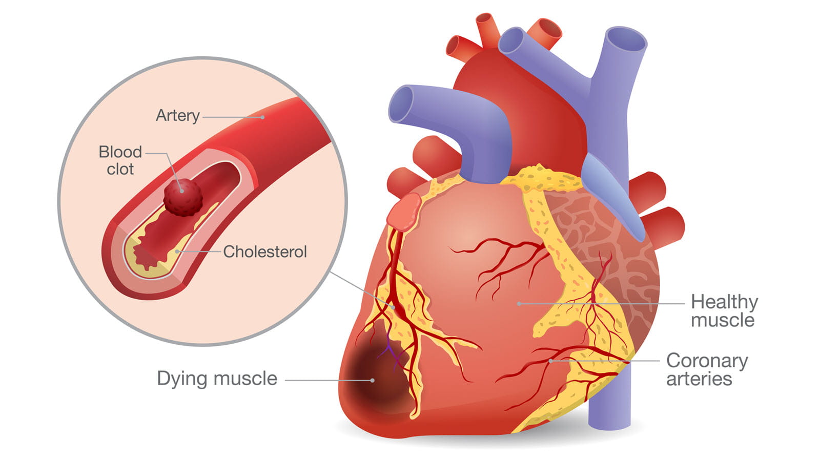
This issue might occur if there is an excessive buildup of fat or cholesterol deposits on the inner wall of the coronary artery, which narrows the artery and feeds blood to the heart. A deposit of fat or cholesterol on the inside wall of an artery is referred to as a plaque. Occasionally, these plaques degrade and create thrombi, which can obstruct capillaries and harm certain cardiac regions. A heart attack, also known as a myocardial infarction, requires emergency care in order to preserve the patient's life.
Risk Factors for a Heart Attack
- Age: The risk of a heart attack is higher in men over 45 and women over 55 than in younger people.
- Usage of tobacco products: Chewing tobacco and its products and smoking cigarettes are also causes of heart attacks.
- High blood pressure or hypertension: A person whose blood pressure is high for a long time is also at risk of a heart attack.
- High triglycerides and cholesterol: The buildup of harmful cholesterol on the inner wall of the arteries causes them to narrow. This type of cholesterol makes it more difficult for blood to pass through the arteries and raises the risk of a heart attack. In a similar vein, elevated blood triglyceride levels raise the risk of myocardial infarction.
- Diabetes: An elevated blood sugar level in the body is a symptom of diabetes. Heart attacks are also more likely as a result of this illness.
- Family history: An individual is more susceptible to heart attacks if there is a history of heart attacks in their family.
- Inadequate exercise and poor diet: Eating meals high in trans fat, salt, and sugar, animal fat, and processed foods, as well as not exercising regularly, raises the chance of having a heart attack.
- Stressful life: An increased risk of heart attack is associated with emotional stress, such as excessive anger or negative thoughts.
- Usage of illicit drugs: Certain behaviors, such as using stimulants like cocaine and amphetamines, have an adverse effect on the coronary artery and raise the risk of a heart attack.
Symptoms of a Heart Attack
Symptoms of a heart attack vary among people. General symptoms of a heart attack are given below:
- Acute chest pain and discomfort that gradually travels to the shoulder, hand, neck, jaw, and even the stomach
- Cold sweats, exhaustion, and dyspnea
- Headache, abrupt lightheadedness, or nausea
- Some people get heart attacks suddenly. However, the majority of patients have warning indications of a heart attack several hours, days, or weeks in advance. These indicators include persistent chest pain or pressure that does not go away even when you relax. These are the early warning signs, and inadequate blood flow to the heart's tissues may be the cause of heart pain.
Preventive Measures
- Keep up a healthy way of living.
- Give up drinking alcohol and stop smoking.
- Maintain a healthy weight.
- Eat a nutritious, well-balanced diet.
- Work out frequently.
- Control your tension.
- High blood pressure and diabetes both increase the risk of having a heart attack. Thus, it is important to control and treat these disorders.
- Go for regular health check-ups.
Diagnosis of Heart Attack and Treatment
The temperature, pulse, and blood pressure are measured in order to diagnose a heart attack. General tests pertaining to the heart, heartbeat, etc. are also part of the diagnosis.
Many methods, such as an electrocardiogram, blood test, echocardiography, CT coronary angiography, and MRI, are used to test for heart attacks.
Angiography
A kind of fluoroscopy X-ray called an angiography is used to check for blood vessel blockages. Blood vessel X-ray pictures are typically not easily observed. But during this procedure, the blood is infused with contrast medium, a unique color. This procedure makes blood flow more visible and makes blood vessel issues more visible. An angiography produces an X-ray picture known as an angiogram.
Numerous cardiac tissues are damaged every minute during a heart attack. Therefore, in order to restore blood flow and oxygen levels, prompt therapy is required. The patient must have an immediate supply of oxygen in order to do this. Particularly, whether there is a partial or total stoppage of blood flow affects how a heart attack is treated. The patient has to be brought to the hospital right away so that the medical staff can treat them and follow their orders. When angioplasty and angiography equipment are unavailable, physicians often begin therapy with blood thinners. In locations where angioplasty and angiography are available, opening the clogged artery requires surgery. Medical procedures like coronary angioplasty and stenting, coronary artery bypass surgery, etc. open blocked arteries.
Coronary Angioplasty and Stenting
Also known as percutaneous coronary intervention (PCI), this medical treatment opens the heart's obstructed coronary artery. A cardiologist performs an angioplasty by inserting a long, thin, flexible tube called a catheter into a hand or leg blood vessel and guiding it to the heart's constricted artery. During angioplasty, a stent—a tiny metal mesh tube—is frequently inserted. In order to preserve regular blood flow, the stent aids in opening constricted or obstructed blood vessels. Because the stent is covered in medication, it opens up blood vessels and keeps them from constricting.
Coronary Artery Bypass Surgery
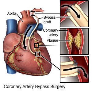
During a heart attack, this emergency surgical procedure—also known as open-heart surgery—is used. Using surgery, a healthy leg blood artery is removed to create a new conduit that will increase blood flow to the heart muscle. Then, a new channel delivers blood that is rich in nutrients and oxygen to the heart tissues.
Heart beat, Pulse rate or Heart rate
The constant and rhythmic contraction and relaxation of the heart muscle produce the sound of a heartbeat. The body's physical state affects the heartbeat. The instrument used to measure the heartbeat is called a stethoscope. A healthy adult's heart beats between 60 and 100 times per minute while they are at rest. This is called the heart rate A slow heartbeat, which is less than 60 times per minute, is called bradycardia, or slow heart. A fast heartbeat, which is more than 100 times per minute, is called tachycardia, or fast heart.
Blood is circulated to different cells and tissues through arteries as a result of the heart muscle's regular, rhythmic contraction and relaxation. Blood pressure on arteries is perceptible from the outside when blood is moving through the artery. We refer to this as the pulse. Heart rate and pulse rate are often identical. Thus, both phrases are interchangeable. The typical pulse rate of a healthy adult is between 60 and 100 beats per minute. One may feel a person's pulse rate by placing fingers on their arm or throat.
Things to remember title
- The heart is the main organ of the circulatory system, responsible for supplying blood to every cell and tissue in the body.
- The heart contains four valves: tricuspid, bicuspid, mitral, and pulmonic.
- Heart attacks are urgent and dangerous situations where the blood supply to the heart tissues is abruptly cut off or obstructed, causing excruciating pain and requiring emergency care.
- Heart attacks can be diagnosed through various tests, including temperature, pulse, blood pressure, and angiography.
- Angiography, a fluoroscopy X-ray, is used to check for blood vessel blockages.
- Coronary angioplasty and stenting, also known as coronary artery bypass surgery, is a medical treatment that opens blocked arteries.
© 2021 Saralmind. All Rights Reserved.
 Login with google