Anatomy and Physiology of the Cardiovascular and Hematologic System
Subject: Medical and Surgical Nursing I (Theory)

Overview
Capillaries, arteries, veins, and the heart make up the cardiovascular system. The removal of carbon dioxide and other waste products from cellular metabolism, delivery of oxygen and other nutrients to the body's tissues, and circulation of blood are the three main duties of the cardiovascular system.
Heart
- The heart is a muscular pump that is housed in the midiastenium. It pumps oxygenated blood into the arterial system and collects deoxygenated blood from the venous system to send it to the lungs where it can be reoxygenated.
- The heart is extends obliquely for 12 to 14 cm (about 5 inches) from the rib to the 5th 2 ^ (nd) intercostals space, the space between the lungs in the thoracic cavity.
- The muscle with the shape of a cone, the heart, rests on the diaphragm. It has a leftward and forward tilt.
- The bottom of the heart is where the apex is located, to the left of the midline. The base is posterior to the sternum and is located at the top of the heart, where the great vessels enter the heart. It measures between 250 and 350 g.
- The pericardium, a sac with two walls, surrounds the heart. The sac that serves to shield the heart from friction is called the pericardium, which is made up of two layers: the serous (inner layer) and fibrous (outer layer) pericardium. Three layers make up the heart. (1) Superficial, covering the surface of the heart, the epicardium merges with the visceral layer of the pericardium. (2) Middle layer of the myocardium, made up of cardiac muscles (3) Endocardium, a slender inner membrane lining the heart chambers.
- The chordae tendinae receive papillary muscles from the endocardial and myocardial surface of the ventricles. The tricuspid and mitral valves' chordae tendinae are connected to them and stop eversion during systole.
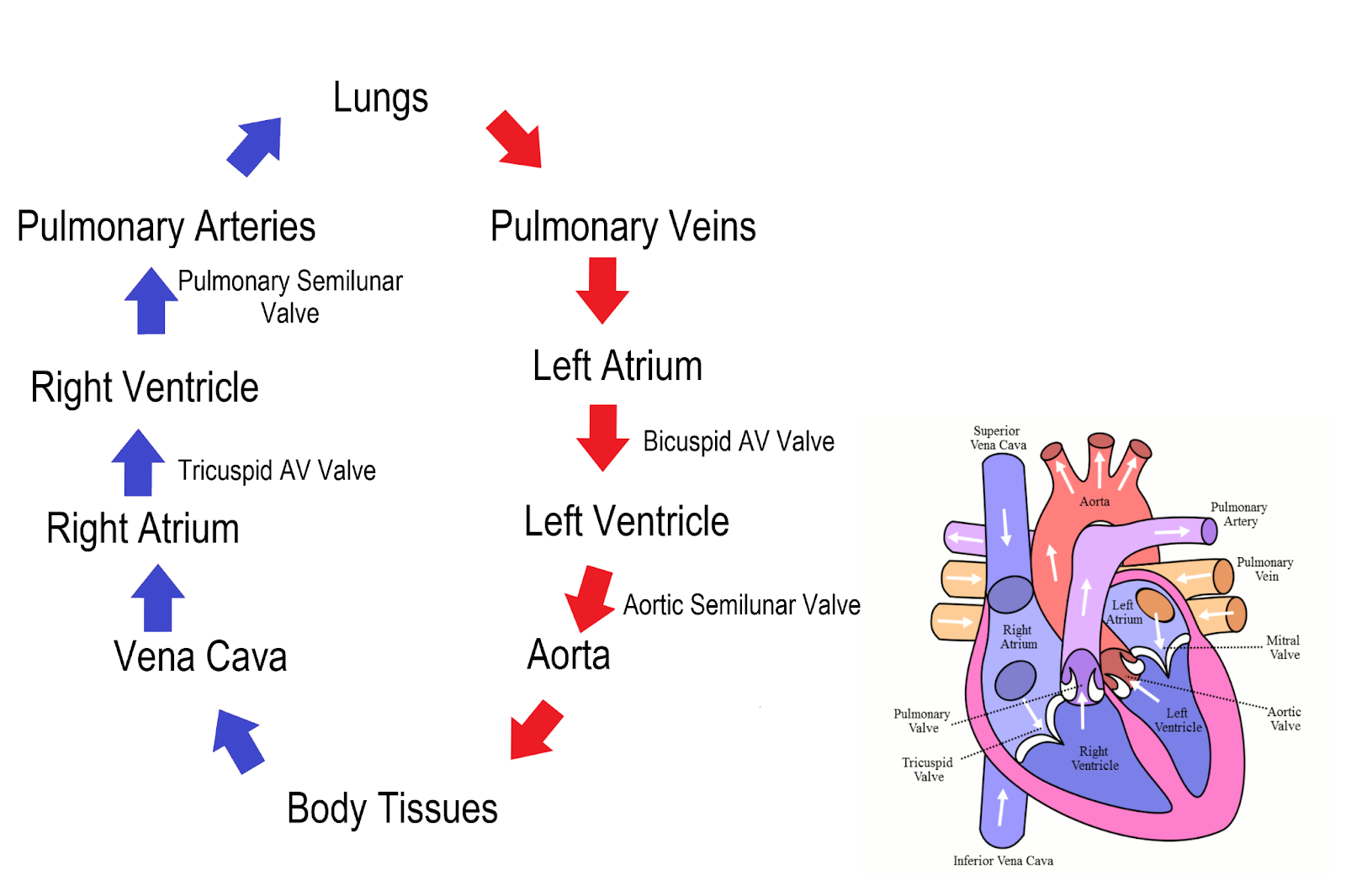
- Each side of the heart has two chambers: a thick-walled bottom pumping chamber and an upper collecting chamber (atrium) (ventricle).
- Through the coronary sinus, inferior vena cava, and superior vena cava, systemic venous blood is delivered to the right atrium. Through the pulmonary veins, the left atrium gets oxygenated blood that is returning to the heart from the lungs.
- When the right ventricle contracts, the pulmonary artery carries deoxygenated blood into the pulmonary circulation. During ventricular systole, the left ventricle pushes blood into the systemic circulation through the aorta.
- Four heart valves—the semi-lunar (pulmonary and aortic) and paired atrioventricular (mitral and tricuspid) valves—control the flow of blood through the heart in a single direction.
- The mitral valve, which has two leaflets attached to the chordate tendinae, is situated between the left atrium and the left ventricle.
- The tricuspid valve, which has three leaflets linked to chordate tendinae, is situated between the right atrium and the right ventricle.
- Permit unidirectional blood flow during ventricular diastole from a specific atrial to a specific ventricle.
- In order to stop reflux flow, valve leaflets open during ventricular diastole and close during ventricular systole.
- first cardiac sound is produced by valve closure (S1). Between the right ventricle and the pulmonary artery is the pulmonary valve.
- Between the left ventricles and the aorta is the aortic valve. During ventricular systole, semilunar valves allow unidirectional blood flow from a particular ventricle to arterial arteries. The valves, which stop reflux blood flow, open when the ventricles contract and close during ventricular diastole. Second heart sound is produced by valve closure.
Blood Supply to the Heart: Coronary Circulation
- The heart muscle needs a plentiful supply of oxygen to meet its own metabolic requirements. The myocardial and conduction system are supplied with blood by the right and left coronary arteries, which branch off the aorta's base slightly above the aortic valve.
- The posterior septal wall, right atrium, right ventricles, sinoatrial node (SA), and atrioventricular node are all perfused by the right coronary artery (RCA) and its branches (AV).
- The left anterior descending (LAD) and circumflex arteries are the two main branches of the left coronary artery (LCA).
- The anterior ventricular septum, the anterior wall of the left ventricle, and the left ventricle's apex are all supplied with blood by the LAD.
- The left atrium, as well as the lateral and posterior surfaces of the left ventricle, receive blood from the circumflex artery. Coronary veins use the coronary sinus to transport blood from the heart back to the right atrium.
- The heart muscle needs a plentiful supply of oxygen to meet its own metabolic requirements.
- The myocardium and conduction system are supplied with blood by the right and left coronary arteries, which branch off the aorta's base (just above the aortic valve).
- The posterior septal wall, right atrium, right ventricles, sinoatrial node (SA), and atrioventricular node are all perfused by the right coronary artery (RCA) and its branches (AV).
- The left anterior descending (LAD) and circumflex arteries are the two main branches of the left coronary artery (LCA).
- While the circumflex artery supplies blood to the left atrium, the lateral, and the posterior surface of the left ventricle, the LAD supplies blood to the anterior wall of the left ventricle, the anterior ventricular septum, and the left ventricle's apex. Coronary veins use the coronary sinus to transport blood from the heart back to the right atrium.
The Conduction System of the Heart
- The heart's conducting system is made up of cardiac muscle cells and conducting fibers, not nerve tissue, which are designed specifically for generating impulses and swiftly transmitting them through the heart. They start the regular cardiac cycle and control the heart's chamber contractions. The mechanism supplies the heart with its regular, automated beat.
- The right atrium's wall, close to the superior vena cava's entrance, houses the Sinoatrial (SA) node, the heart's main pacemaker.
- The AV node card, which is situated in the lower right atrium next to the interatrial septum, originates the impulse (60–100b/m).
- There is no pacemaker in the nodal tissue itself. Its surrounding tissue, known as the junctional tissue, has pacemaker cells that can naturally beat at a rate of 40. 60 beats per minute.
- The A node gives rise to the bundle of his, which sends an impulse to the bundle branch system. Purkinje Fibers penetrate deeply into the myocardium from the bundle branches in the endocardium.
- The muscle receives impulses quickly to help with depolarization and contraction from the purkinje fibers, which discharge at an inherent rate of 20.40 beats per minute or even more slowly.
- The cardia muscle's electrophysiologic characteristics control heart rate and rhythm.
- One of the characteristics is automaticity, or the capacity to produce and release an electrical impulse on demand. 2) Excitability: A cell's capacity to react to an electrical stimulus. (3) conductivity, which is the capacity of one cell to transmit an electrical impulse to another; (4) contractility, which is the capacity of the cell to shorten and lengthen its fibers (5) During the entire systole and early diastole, the cardiac muscle is refractory and cannot be released. This time frame prevents tetanus and the summing of waves.
- The series of events that take place as the heart beats is known as the cardiac cycle. The cardiac cycle's event sequence is split into two phases: (1) The heart fills with blood during the diastole phase, and (2) the heart contracts during the systole phase to pump blood to the arteries. Blood is pumped through the body's pulmonary and systemic circuits as the heart beats.
Things to remember title
© 2021 Saralmind. All Rights Reserved.
 Login with google